

Loading, please wait...
Loading, please wait...

Aneurysmal subarachnoid hemorrhage remains one of the most perilous neurosurgical emergencies worldwide, primarily because secondary neurovascular complications can drastically derail patient recovery. Following initial aneurysm securement, clinicians frequently encounter delayed cerebral ischemia and arterial narrowing that impair microvascular perfusion. Clinicians have traditionally relied on induced hypertension, hypervolemia, and rescue endovascular interventions to counteract secondary ischemic deterioration. However, these conventional modalities carry profound risks, including pulmonary edema, myocardial strain, and arterial dissection. Consequently, neurocritical care teams have increasingly evaluated intravenous milrinone as an early, targeted hemodynamic strategy to resolve vascular narrowing and optimize cerebral microcirculation without demanding hazardous systemic pressures.
Cerebral vasospasm and delayed ischemia develop through a complex cascade initiated by subarachnoid blood breakdown products. Oxyhemoglobin degrades and triggers extensive endothelial dysfunction, robust oxidative stress, and the depletion of endogenous nitric oxide. Consequently, intracellular cyclic adenosine monophosphate levels drop inside vascular smooth muscle, provoking persistent microvascular and macrovascular constriction. Here, intravenous milrinone operates as a selective phosphodiesterase-3 inhibitor that blocks the breakdown of cyclic adenosine monophosphate. Therefore, the drug promotes smooth muscle relaxation and prompt luminal vasodilation across cerebral vessels.
In addition to direct luminal dilatation, milrinone exerts meaningful inotropic and lusitropic effects on the myocardium. Thus, the drug enhances cardiac output and forward stroke volume without excessively escalating myocardial oxygen consumption. Furthermore, emerging translational evidence suggests that phosphodiesterase-3 inhibition mitigates microvascular thrombosis and blunts local inflammatory cascades within the subarachnoid space. Because delayed cerebral ischemia involves cortical spreading depolarization and microcirculatory thrombosis beyond visible angiographic spasm, this multifaceted pharmacodynamic profile delivers comprehensive neuroprotection. As a result, starting intravenous therapy early addresses both upstream conduit spasm and downstream microcirculatory failure.
Recent observational cohort data provide compelling evidence regarding the utility of early intravenous milrinone in aneurysmal subarachnoid hemorrhage. In a landmark retrospective investigation evaluating 336 patients, 130 individuals developed confirmed cerebral vasospasm or delayed cerebral ischemia. Among the patients presenting with overt neurological deterioration, 59 percent demonstrated rapid clinical improvement after clinicians initiated intravenous milrinone. Therefore, the infusion served as an effective pharmacological rescue for neurological deficits.
Equally important, early initiation dramatically curtails the need for aggressive rescue treatments. In this clinical evaluation, only 20 percent of patients in the milrinone group required escalation to vasopressor support. In sharp contrast, 84 percent of patients in the non-milrinone cohort required vasopressor infusions to sustain induced hypertension. Furthermore, the milrinone cohort showed a significantly lower rate of invasive rescue endovascular therapy. By establishing stable cerebral perfusion through active vasodilation and increased cardiac index, milrinone eliminates the need for extreme hypertensive surges. Consequently, intensive care units observe fewer cardiovascular complications and lower nursing burdens related to invasive arterial line titration.
Neurointensivists generally implement intravenous milrinone through structured, step-wise institutional protocols. Clinicians usually omit the standard cardiac loading bolus to avoid acute systemic hypotension. Instead, teams start a continuous maintenance infusion ranging between 0.25 and 0.75 micrograms per kilogram per minute. Thereafter, bedside intensivists titrate the infusion rate according to daily transcranial Doppler velocities, serial neurological evaluations, and multimodal perfusion imaging findings.
Simultaneously, practitioners maintain strict euvolemia and closely monitor invasive arterial blood pressure. Because milrinone induces mild systemic peripheral vasodilation, clinicians occasionally co-administer low-dose norepinephrine to preserve adequate cerebral perfusion pressure. Moreover, continuous cardiac telemetry remains mandatory throughout the entire infusion course. Clinicians should proactively observe for atrial or ventricular arrhythmias and baseline corrected QT interval prolongation. Additionally, physicians routinely monitor daily serum potassium and magnesium levels, as hypokalemia increases arrhythmogenic susceptibility during phosphodiesterase inhibition. When patients demonstrate sustained clinical stability for 48 to 72 hours beyond the high-risk vasospasm window, teams taper the infusion gradually to prevent rebound vasospasm.
Although early intravenous milrinone offers undeniable therapeutic value, neurocritical care teams must navigate several practical safety considerations. First, peripheral vasodilation may provoke refractory systemic hypotension, particularly in volume-depleted intensive care patients. Therefore, clinicians must achieve euvolemia before drug initiation and maintain invasive hemodynamic monitoring throughout therapy. Second, milrinone undergoes extensive renal elimination. Consequently, intensive care specialists must adjust dosage regimens in patients suffering from acute kidney injury or pre-existing renal insufficiency to prevent drug accumulation.
Third, milrinone therapy frequently induces sustained polyuria through altered renal hemodynamics and natriuresis. Bedside nurses must measure hourly fluid balance accurately to avert sudden hypovolemia, which could otherwise worsen cerebral hypoperfusion. Fourth, drug-induced thrombocytopenia and mild electrolyte wasting demand vigilant laboratory surveillance. Fortunately, clinical experience confirms that serious adverse reactions rarely require permanent drug discontinuation when clinicians utilize continuous telemetry and standardized titration algorithms. Nevertheless, clinicians must carefully weigh individual cardiovascular risk profiles before deciding between intravenous inodilator infusion and conventional induced hypertension.
The neurointensive care community is actively transitioning away from non-selective systemic induced hypertension toward personalized, mechanistically guided neurovascular care. Historical strategies that relied heavily on aggressive hypervolemia and severe induced hypertension frequently caused cardiogenic shock, flash pulmonary edema, and hemorrhagic infarctions. Conversely, early intravenous milrinone provides a balanced alternative that improves microvascular oxygen delivery without threatening systemic capillary beds.
Nevertheless, clinicians must recognize the current evidence limitations. Most supporting literature consists of retrospective analyses, non-randomized before-and-after studies, and single-center institutional registries. Therefore, neurosurgeons and intensivists eagerly await results from prospective, randomized controlled clinical trials to define exact dosing windows, long-term functional recovery, and patient stratification biomarkers. Until large-scale trial findings emerge, multi-professional intensive care units should establish clear protocols for early identification of vasospasm, timely infusion initiation, and proactive electrolyte stabilization. Adopting such standardized frameworks ensures optimal cerebral salvage and enhances overall patient recovery.
Intravenous milrinone acts as a phosphodiesterase-3 inhibitor that raises intracellular cyclic adenosine monophosphate in vascular smooth muscle and myocardium. Consequently, it promotes potent cerebral arterial vasodilation, increases cardiac contractility, enhances microcirculatory cerebral blood flow, and suppresses local vascular inflammation without demanding hazardous levels of induced systemic hypertension.
Early intravenous milrinone substantially lowers the necessity for adjunctive vasopressor therapy. By improving forward cardiac output and dilating cerebral vessels, only twenty percent of patients require vasopressors compared to over eighty percent managed conventionally. Furthermore, this physiological optimization significantly reduces the requirement for invasive endovascular balloon or chemical angioplasty.
Clinicians must maintain continuous invasive arterial blood pressure monitoring and cardiac telemetry to promptly identify hypotension or arrhythmias. Additionally, neurocritical teams must monitor renal function, track platelet counts, replace urinary electrolyte losses, and perform daily transcranial Doppler ultrasonography to guide titration and evaluate therapeutic response accurately.
Disclaimer: This content is for informational and educational purposes only... Refer to the latest local and national guidelines for clinical practice.
References
1. Baang HY et al. Treatment Effect of Early Intravenous Milrinone for Cerebral Vasospasm or Delayed Cerebral Ischemia After Aneurysmal Subarachnoid Hemorrhage. Neurocrit Care. 2025 Aug. doi: 10.1007/s12028-025-02260-x. PMID: 40329063.
2. Lannes M, Teitelbaum J, del Pilar Murias Quirós M, et al. Milrinone for cerebral vasospasm in subarachnoid hemorrhage: the MILRISPASM controlled before-after study. Neurocrit Care. 2020;33(2):505-514.
3. Bernier TD, Schontz MJ, Izzy S, et al. Treatment of Subarachnoid Hemorrhage-associated Delayed Cerebral Ischemia with Milrinone: A Review and Proposal. Neurocrit Care. 2021;35(2):541-551.

Read summarized clinical updates, watch expert medical content, and earn CME certifications right from your smartphone.
Open your smartphone camera to scan and install.





Early intravenous milrinone therapy demonstrates notable efficacy in reversing cerebral vasospasm and mitigating delayed cerebral ischemia after aneurysmal subarachnoid hemorrhage, significantly curtailing vasopressor dependency and rescue interventions.
Today

Standardizing continuous physiological waveforms in EHRs has long challenged clinical teams. The novel TIDE framework leverages HL7 FHIR R4 to decouple queryable clinical metadata from massive raw signal repositories, unlocking scalable telemetry exchange for bedside care and predictive research.
Today

A secondary analysis of the MOMS clinical trial reveals that infant sex does not influence neonatal opioid withdrawal syndrome severity in buprenorphine-exposed newborns. Maternal co-exposures and clinical factors drive outcomes, highlighting the need for objective assessment and supportive breastfeeding.
Today
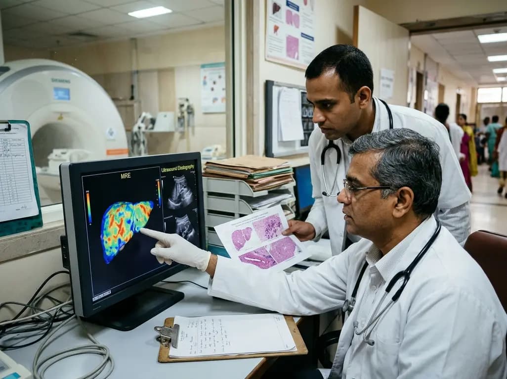
A comparative study reveals magnetic resonance elastography outperforms ultrasound elastography in assessing liver fibrosis in Fontan-associated liver disease, demonstrating stronger histological agreement and superior discrimination.
Today

A systematic review and meta-analysis highlights the substantial impact of plyometric jump training on jump performance, reactive strength, and landing kinematics in female team-sport athletes, providing evidence-based programming guidelines for sports medicine and orthopedics.
Today

A comprehensive review of the role of growth hormone in diabetic kidney disease and podocytopathy. Learn how elevated GH activates ZEB2, Notch, and TGF-beta1, driving podocyte loss and glomerulosclerosis, and explore novel targeted therapeutic approaches.
Today
