.webp&w=384&q=75)

Loading, please wait...
Loading, please wait...
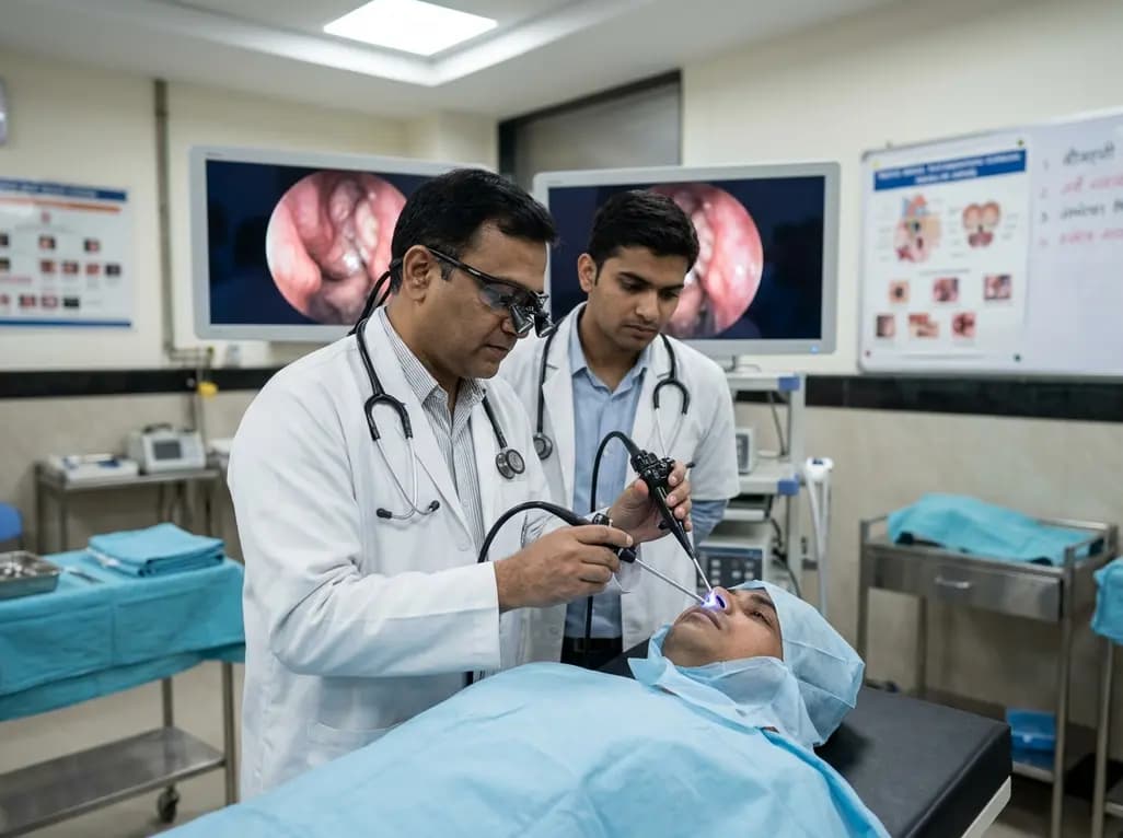
For decades, neurosurgeons and otolaryngologists traditionally favored conservative observation for managing traumatic skull base dural disruption. Historically, open craniotomies carried significant surgical morbidity, which justified a watchful waiting strategy. However, modern endonasal techniques now allow surgeon teams to access complex cranial base anatomy with minimal tissue disruption. Emerging clinical evidence shows that early endoscopic skull base repair provides exceptional therapeutic closure while preventing severe secondary infectious complications.
A landmark retrospective study evaluated 385 patients with documented traumatic dural disruption. The study investigated surgical efficacy alongside the clinical significance of indirect radiographic markers like pneumocephalus. Interestingly, the research demonstrated that conservative management often led to preventable intracranial infections. Consequently, clinicians are now reconsidering traditional observation protocols in favor of early endonasal intervention.
Historically, clinicians managed traumatic dural defects conservatively because transcranial operative repair carried substantial surgical risks. Practitioners often prescribed strict bed rest, head elevation, and lumbar drains while waiting for spontaneous closure. However, conservative observation exposes patients to prolonged hospital stays and severe ascending central nervous system infections. Furthermore, persistent dural defects create a direct conduit between the unsterile sinonasal cavity and the sterile subarachnoid space.
In the landmark retrospective cohort of 385 trauma patients, 102 individuals underwent initial conservative management. Remarkably, 34 of these patients, representing 33.3 percent, subsequently developed meningitis or intracerebral infections. This high complication rate highlights the primary vulnerability of conservative protocols. In addition, delayed surgical repair often occurs in the setting of active central nervous system inflammation, which increases operative complexity. Therefore, relying solely on observation frequently converts a manageable structural injury into a life-threatening infectious emergency. Early definitive intervention effectively eliminates this prolonged window of vulnerability, protecting patients from severe intracranial sequelae.
Identifying dural disruption quickly remains a critical priority in acute trauma evaluation. Traditionally, overt cerebrospinal fluid rhinorrhea served as the primary trigger for surgical repair. However, relying exclusively on active fluid discharge can lead to missed diagnoses because physical examination findings are often subtle or intermittent. In the study cohort, 219 patients presented with frank cerebrospinal fluid rhinorrhea, whereas 254 patients demonstrated radiographic pneumocephalus.
Importantly, pneumocephalus represents clear evidence of structural dural violation, even when overt cerebrospinal fluid leaking is absent. Intracranial air indicates that atmospheric pressure from the sinonasal tract has breached the dural barrier. Consequently, surgeons must recognize pneumocephalus as an independent marker of dural disruption that justifies early intervention. Utilizing comprehensive high-resolution computed tomography allows clinicians to identify small bone fragments and occult air collections rapidly. Therefore, incorporating endoscopic skull base repair early in the clinical pathway addresses structural defects before infectious pathogens migrate into intracranial compartments.
Transnasal endoscopic reconstruction offers unparalleled direct visualization and meticulous tissue handling without external skin incisions. In the evaluated study, primary endoscopic closure achieved an extraordinary success rate of 97.4 percent, repairing 375 out of 385 patients during the initial procedure. Furthermore, secondary endoscopic revision increased the overall definitive success rate to 99.0 percent. These remarkable outcomes confirm that endonasal repair delivers durable long-term structural integrity.
The anatomical distribution of traumatic skull base defects varied across distinct sinonasal regions. The ethmoid sinuses represented the most frequent site of involvement with 244 cases, followed closely by the frontal sinuses with 212 cases and the sphenoid sinuses with 157 cases. Many patients presented with complex fractures that involved multiple contiguous anatomical zones. Notably, patients requiring multiple surgical procedures were predominantly those who suffered penetrating head trauma rather than blunt impacts. Multilayer closure techniques, utilizing vascularized nasoseptal flaps, fascia lata, or free mucosal grafts, provided robust seals across these diverse anatomical regions.
The development of intracranial infection represents the most devastating outcome following traumatic skull base dural disruption. Bacteria from the nasal passages can rapidly colonize subarachnoid spaces, causing meningitis, brain abscesses, or subdural empyema. These infectious complications carry high mortality rates and frequently result in permanent neurological deficits. The retrospective data clearly demonstrated that observation-based algorithms carry substantial infectious risk that practitioners can avoid through prompt surgical repair.
Moreover, conservative management often requires prolonged bed rest, which increases systemic risks such as deep vein thrombosis, pulmonary embolism, and physical deconditioning. Extended hospitalizations also impose significant financial costs on healthcare systems and patients alike. By contrast, early endonasal intervention accelerates clinical recovery, shortens intensive care unit stays, and minimizes overall hospital length of stay. Consequently, early repair provides both superior clinical safety and substantial economic advantages compared to conservative watchful waiting protocols.
Optimizing clinical outcomes for complex skull base trauma requires coordinated multidisciplinary care. Otolaryngologists, neurosurgeons, radiographers, and critical care specialists must collaborate closely during early trauma resuscitation. Advanced image-guided navigation systems allow surgical teams to localize small fractures precisely and navigate distorted anatomy safely. In addition, vascularized tissue transfer techniques have revolutionized skull base reconstruction by providing rapid healing and excellent infection resistance.
Future trauma protocols should systematically incorporate early endonasal surgical evaluation for all patients presenting with radiographic pneumocephalus or documented skull base fractures. Relying on conservative management should no longer remain the default therapeutic paradigm. Instead, clinical centers should adopt early repair strategies to prevent preventable intracranial infections and improve long-term functional recovery. As endoscopic technology and intraoperative imaging continue to advance, minimally invasive skull base reconstruction will solidify its role as the definitive standard of care in modern trauma management.
Pneumocephalus indicates a direct mechanical breach between the unsterile sinonasal cavity and the sterile subarachnoid space. Even without active cerebrospinal fluid leakage, trapped intracranial air confirms definite dural disruption. Early endoscopic repair seals this mechanical opening promptly, preventing pathogenic sinus bacteria from migrating into the central nervous system and causing life-threatening infections like bacterial meningitis or intracerebral abscesses. Consequently, identifying pneumocephalus justifies early surgical intervention.
Primary endoscopic repair is exceptionally successful for traumatic dural injuries, achieving a primary closure success rate of 97.4 percent in recent large clinical studies. When minor endoscopic revision surgery is performed for persistent defects, the overall cumulative success rate reaches 99.0 percent. This minimally invasive technique provides robust, durable dural closure without requiring traditional craniotomy incisions, thereby significantly reducing surgical morbidity and patient recovery times.
Conservative observation carries a significant risk of ascending intracranial infection. Research indicates that over 33 percent of patients managed conservatively subsequently develop severe meningitis or intracerebral infections. Furthermore, conservative strategies often involve prolonged bed rest, which increases systemic complications like deep vein thrombosis, pulmonary embolism, extended hospitalization costs, and delayed overall functional recovery. Therefore, early surgical intervention provides a much safer clinical alternative.
Disclaimer: This content is for informational and educational purposes only and does not constitute medical advice, diagnosis, or treatment. Always seek the advice of a qualified healthcare provider with any questions you may have regarding a medical condition. Refer to the latest local and national guidelines for clinical practice.
References
Grayson JW et al. Early Endoscopic Repair of Traumatic Skull Base Dural Injuries: Challenging the Dogma of Conservative Management. Int Forum Allergy Rhinol. 2026 Aug 11. doi: 10.1002/alr.70238. PMID: 42579312.
Govindaraj S, Del Signore AG, Kennedy DW. Endoscopic Skull Base Defect Repair. Otolaryngol Clin North Am. 2016;49(1):141-152.

Read summarized clinical updates, watch expert medical content, and earn CME certifications right from your smartphone.
Open your smartphone camera to scan and install.





A recent landmark study demonstrates that early endoscopic repair of traumatic skull base dural injuries achieves a 99% overall success rate while significantly reducing infectious complications compared to conservative observation, challenging traditional clinical management algorithms.
last month
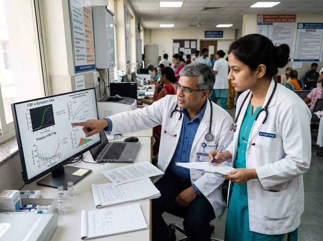
A new PPMI cohort study demonstrates that combining blood microRNA-7-5p with CSF alpha-synuclein seed amplification assay significantly improves the prediction of Parkinson's disease phenoconversion, offering clinicians and researchers a powerful tool for patient risk stratification and clinical trial enrichment.
Today
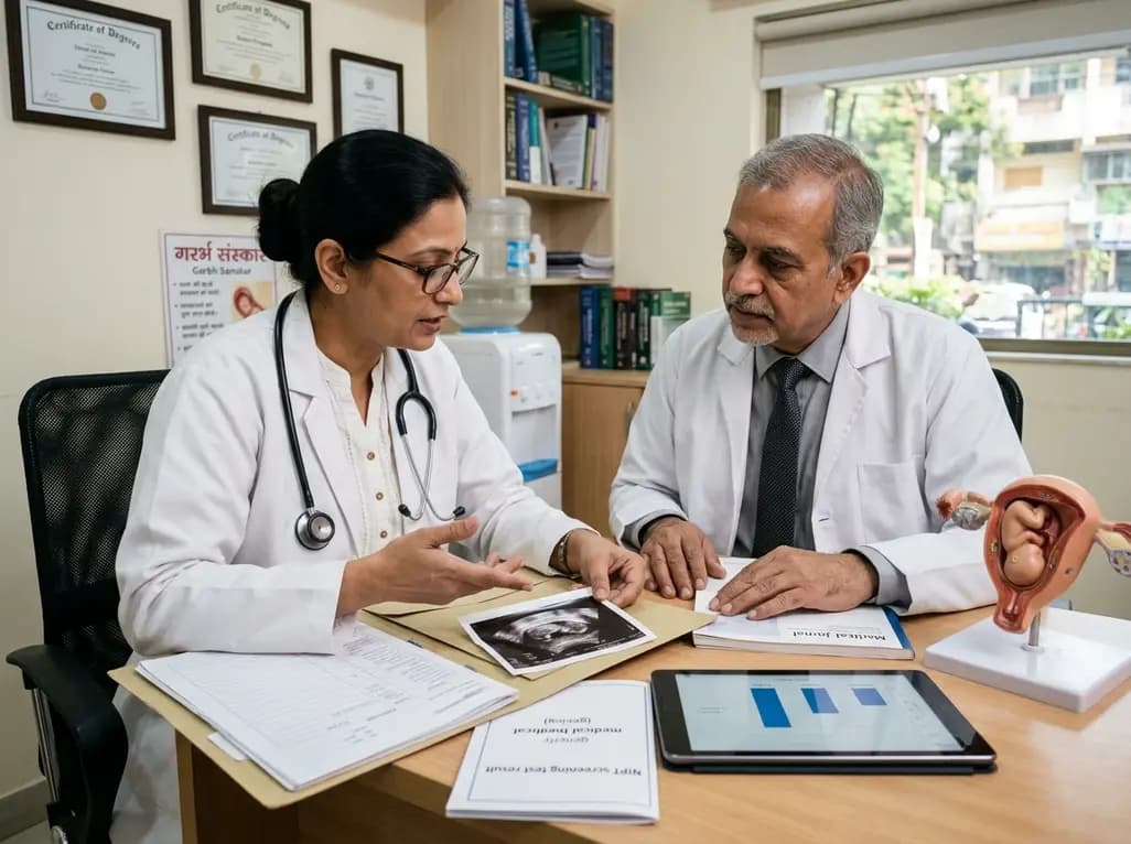
A large retrospective study evaluates the screening accuracy, positive predictive value, and test failure rates of noninvasive prenatal testing (NIPT) in assisted reproductive technology (ART) pregnancies compared to spontaneous conceptions, highlighting key considerations for pretest counseling.
Today

Differential oxygenation during peripheral TA-NRP can cause undetected coronary hypoxemia due to arch vessel clamping. A novel circuit with continuous aortic root monitoring detects desaturation early, facilitating left ventricular unloading and preventing allograft dysfunction and unnecessary heart discard.
Today

A randomized controlled trial compared ultrasound-guided intra-articular injections of PRP, hydrolyzed type I collagen, and hyaluronic acid in knee osteoarthritis. All therapies demonstrated significant improvements in WOMAC and pain scores at six months, supporting hydrolyzed collagen as a viable biological option.
Today

A scoping review evaluates 116 studies on AI in women's reproductive health, finding that 83.6% carry high risk of bias. While ensemble models show promise, disparities and lack of validation hinder LMIC deployment.
Today
