

Loading, please wait...
Loading, please wait...
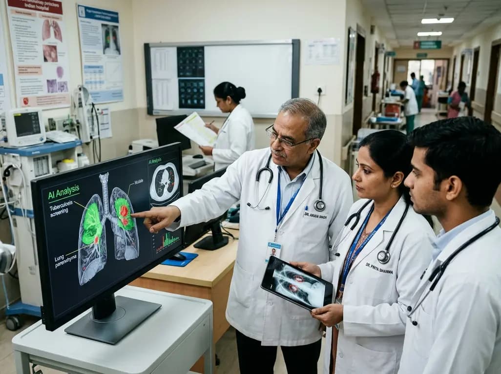
At NAPCON 2026 in Jaipur, respiratory physicians gathered to discuss transformative clinical technologies. Specifically, artificial intelligence now offers unprecedented support for the early detection of TB and lung cancer across India. Leading experts emphasized that deep learning models can rapidly analyze diagnostic images. Consequently, these algorithms assist clinicians in triaging high-risk patients who require urgent intervention. As diagnostic demands escalate nationwide, integrating software solutions into public health frameworks has become a top clinical priority.
India accounts for approximately 26 percent of the global tuberculosis caseload. Therefore, primary care physicians face persistent diagnostic delays, especially in peripheral communities. Early detection of TB remains essential to halt disease transmission and reduce pulmonary morbidity. When healthcare workers employ computer-aided detection software on digital chest X-rays, they identify suspicious radiographic lesions within minutes. Consequently, mobile screening vans can evaluate hundreds of individuals in remote districts each day. Furthermore, automated image analysis highlights apical infiltrates, cavitary lesions, and subtle consolidation that human readers might overlook during rapid mass triage. Dr. Randeep Guleria highlighted that these automated tools streamline clinical decision-making. As a result, frontline doctors can direct high-risk individuals toward rapid molecular microbiological testing without delay. In addition, early intervention prevents secondary household transmission and curtails lung parenchymal damage. However, clinicians must recognize that artificial intelligence operates solely as a preliminary triage filter. It directs symptomatic individuals toward rapid sputum assays such as Truenat or GeneXpert. Ultimately, integrating artificial intelligence into community screening pathways bridges longstanding geographic disparities and optimizes resource allocation across regional healthcare centers. Specifically, public health authorities can deploy these digital solutions across rural wellness centers to strengthen universal diagnostic coverage.
The World Health Organization explicitly endorses computer-aided detection software as an alternative to human radiograph interpretation for tuberculosis screening. However, Dr. Raj Kumar from the Vallabhbhai Patel Chest Institute emphasized that algorithms cannot replace clinical acumen. Instead, medical teams must integrate artificial intelligence into established diagnostic algorithms. First, digital radiography units capture posteroanterior chest views in outpatient clinics. Next, calibrated algorithms generate an automated abnormality score based on pre-trained neural networks. If the score exceeds the validated threshold, the clinical team immediately orders confirmatory microbiological tests. Consequently, this triage mechanism conserves expensive molecular reagents for individuals with demonstrable radiographic pathology. Moreover, automated screening reduces the workload of overburdened radiologists in government hospitals. Meanwhile, general practitioners receive immediate notifications regarding abnormal findings, which eliminates weeks of referral waiting time. In addition, software platforms can flag concurrent abnormalities like cardiomegaly or pleural effusions. Therefore, structured integration safeguards patient safety while accelerating disease confirmation. Ultimately, successful adoption requires robust electronic medical record systems and standard operating protocols that ensure seamless communication between diagnostic hubs and peripheral clinics. Furthermore, rigorous quality control checks prevent false-positive spikes from distorting diagnostic pathways in busy regional laboratories.
Lung cancer remains one of the deadliest malignancies worldwide because clinicians frequently diagnose patients at advanced stages. At NAPCON 2026, Dr. Mohammed Munavvar emphasized that patient prognosis depends directly on early tumor detection. Low-dose computed tomography can identify small malignant nodules before symptoms appear. However, visual interpretation of volumetric CT scans demands substantial radiologic time and expertise. Artificial intelligence significantly alleviates this clinical bottleneck. Specifically, deep learning architectures examine thin-slice chest scans to detect sub-centimeter pulmonary nodules. Furthermore, modern computer algorithms measure nodule volume, calculate doubling times, and assess margins for spiculation. Consequently, interventional pulmonologists can accurately stratify nodules using standardized Lung-RADS criteria. In addition, machine learning models analyze subtle textural features within ground-glass opacities that indicate early-stage adenocarcinoma. As a result, multidisciplinary oncology teams can schedule prompt biopsies or thoracic resections for high-risk lesions. Conversely, low-risk solid nodules can undergo conservative serial imaging, which spares patients from unnecessary invasive procedures. Thus, automated nodule assessment empowers chest specialists to detect thoracic malignancies during resectable stages, significantly improving overall survival rates. Meanwhile, early diagnostic intervention minimizes surgical morbidity and substantially lowers the overall financial toxicity of protracted oncologic therapies.
Deploying artificial intelligence across India’s diverse healthcare ecosystem requires addressing several operational hurdles. First, algorithm developers must validate diagnostic tools on native Indian demographic datasets. Many commercial models trained exclusively on Western populations perform unpredictably when confronted with endemic conditions like fibrocavitary tuberculosis scars or occupational pneumoconiosis. Therefore, domestic clinical trials remain vital to establish precise diagnostic specificity and sensitivity. Second, rural health clinics often lack reliable electrical power, high-speed internet connectivity, and advanced computing hardware. Consequently, technology partners must engineer lightweight edge-AI applications that function offline on compact, battery-powered radiographic machinery. Furthermore, public health administrators must ensure affordable pricing for public health procurement. High proprietary software licensing fees could otherwise widen existing healthcare disparities between metropolitan private centers and underfunded rural clinics. In addition, regulatory bodies must formulate clear standards for software safety, data privacy, and algorithm updates. Notably, healthcare administrators must establish rigorous audit pathways to detect automated diagnostic drift over time. Ultimately, solving these logistical, infrastructural, and regulatory challenges will allow India to unlock the full clinical potential of digital screening technologies across all administrative districts. Accordingly, coordinated multistakeholder partnerships remain critical to ensure sustainable, widespread community deployment.
Although artificial intelligence delivers impressive analytic capability, it cannot replace bedside clinical judgment. Pulmonologists at NAPCON 2026 unanimously affirmed that automated software serves strictly as an assistive tool. For instance, computer vision can highlight an abnormal pulmonary opacity, but it cannot conduct a physical examination or elicit an occupational history. Similarly, algorithms cannot weigh complex comorbid states like immunocompromise, diabetes mellitus, or chronic kidney disease during therapeutic decision-making. Therefore, chest physicians must interpret AI recommendations within the comprehensive clinical context of each patient. Moreover, medical educators must reform pulmonology residency curricula to incorporate digital literacy and algorithmic interpretation. Trainees must understand statistical performance metrics, such as positive predictive value and false discovery rates, to critically appraise AI outputs. Furthermore, clinicians must maintain active vigilance against automation bias, where doctors uncritically accept software conclusions without verification. Ultimately, ethical medical practice requires treating physicians to hold full accountability for diagnostic and therapeutic actions. By combining computational speed with nuanced clinical empathy, pulmonologists can elevate diagnostic standards while delivering compassionate, evidence-based pulmonary care to all patients. In summary, modern technology amplifies diagnostic precision while seasoned physician judgment protects patient well-being at the bedside.
Q1: How does computer-aided detection software improve tuberculosis screening in resource-limited settings?
Computer-aided detection software rapidly analyzes digital chest radiographs to identify subtle apical infiltrates and consolidations indicative of tuberculosis. In underserved areas lacking onsite radiologists, this automated triage tool immediately flags individuals with high abnormality scores. Consequently, local healthcare workers can promptly collect sputum samples for confirmatory molecular testing. Furthermore, this streamlined approach minimizes diagnostic delays, reduces unnecessary travel for rural patients, and conserves valuable healthcare resources.
Q2: What role does artificial intelligence play in evaluating pulmonary nodules for lung cancer?
Artificial intelligence algorithms inspect volumetric computed tomography scans to detect sub-centimeter pulmonary nodules that might escape human detection. In addition, these deep learning tools automatically calculate nodule dimensions, doubling times, and morphological features like border spiculation. Consequently, interventional pulmonologists can accurately categorize lesions using standardized Lung-RADS criteria. Therefore, clinicians can schedule timely biopsies for suspicious malignant nodules while recommending conservative follow-up imaging for low-risk findings, preventing invasive procedures.
Q3: Why is microbiological confirmation essential after an AI tool flags a chest radiograph?
Artificial intelligence serves strictly as a sensitive screening mechanism rather than a definitive diagnostic test. Notably, benign conditions like healed fibrocalcific scars, bronchiectasis, fungal infections, or residual inflammatory changes can generate false-positive radiographic alerts. Therefore, clinicians must verify every positive screening result using microbiological assays such as sputum smear microscopy, culture, or rapid molecular tests. Sputum confirmation ensures precise bacteriological identification and antimicrobial susceptibility profiling before clinicians initiate anti-tubercular therapy.
Disclaimer: This content is for informational and educational purposes only. It does not constitute medical advice or replace professional judgment. Refer to the latest local and national guidelines for clinical practice.
References

Read summarized clinical updates, watch expert medical content, and earn CME certifications right from your smartphone.
Open your smartphone camera to scan and install.





At NAPCON 2026, leading chest physicians highlighted the transformative role of artificial intelligence in screening for tuberculosis and malignant pulmonary nodules. By triaging high-risk chest radiographs and low-dose CT scans, AI aids timely clinical decisions while confirmatory testing remains mandatory.
Today
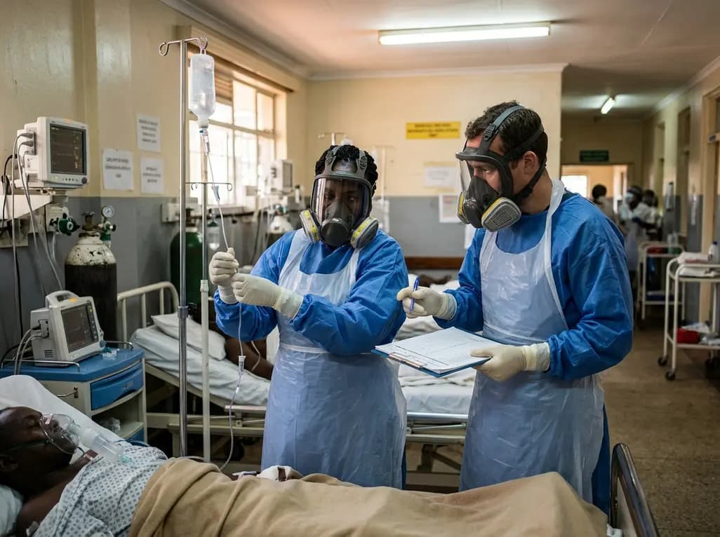
Health authorities in Kenya have confirmed two fatalities among nine cases of Ebola linked to regional transmission. Clinicians worldwide must maintain heightened vigilance, screen international travelers promptly, enforce strict infection prevention, and deliver immediate supportive care for suspected cases.
Today
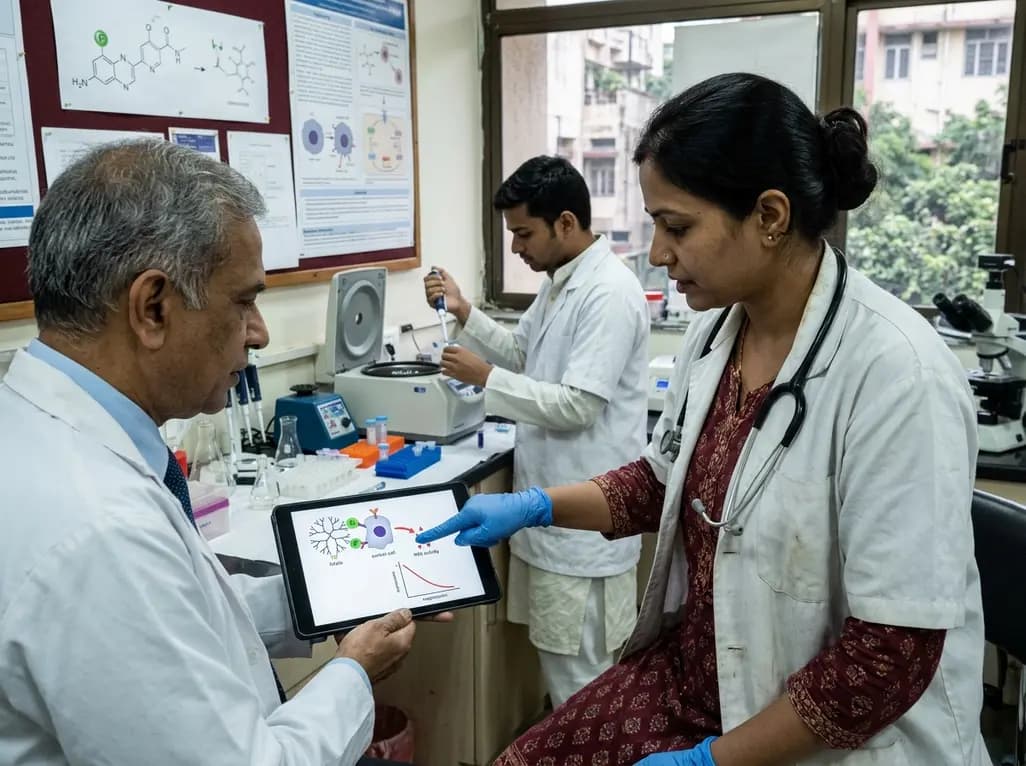
A novel folate-conjugated tecto-dendrimer nanoplatform successfully delivers siRNA to achieve targeted HK2 silencing, disrupting ovarian cancer glycolysis and suppressing lactate output.
Today

A comparative non-contact study reveals that high-power radiofrequency delivery with the QDOT MICRO catheter induces an artifactual contact force rise. Electrophysiologists must understand this power-dependent thermal and electrical phenomenon to avoid inadvertent catheter withdrawal and ineffective ablation.
Today
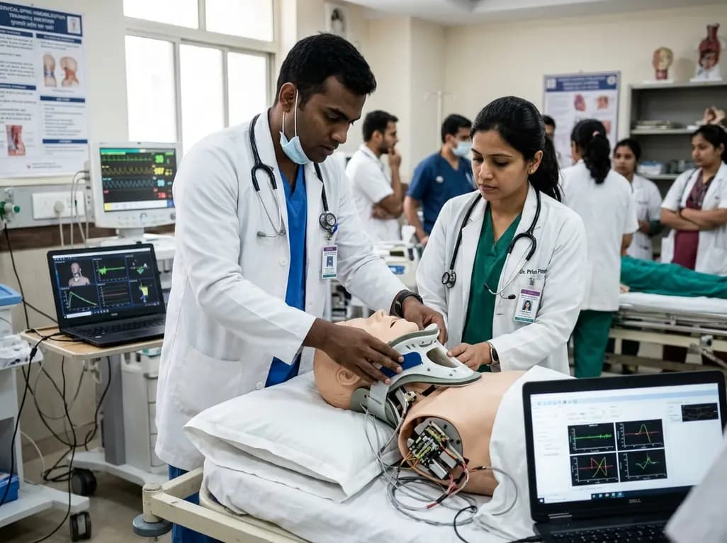
A novel proof-of-concept study presents a six-degree-of-freedom articulated manikin neck equipped with linear potentiometers. The system provides real-time kinematic motion feedback during cervical immobilisation, delivering sub-degree tracking accuracy to improve pre-hospital trauma training and patient safety.
Today
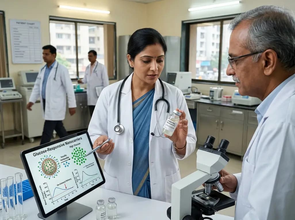
Novel glucose-responsive mPEG-PSA nanocarriers deliver insulin aspart on demand, achieving dual postprandial and basal glycemic control without hypoglycemia in diabetic models. This biomaterial breakthrough offers promising clinical implications for physiologic diabetes management.
Today
