

Loading, please wait...
Loading, please wait...

Severe traumatic brain injury remains a formidable surgical emergency across intensive care units and trauma centers worldwide. When surgeons perform an emergency operation for mass lesions, dynamic craniotomy has emerged as an innovative reconstructive strategy designed to alleviate life-threatening intracranial pressure. Traditionally, neurosurgeons face a difficult dilemma during surgical evacuation of acute hematomas. If they rigidly secure the bone flap with traditional fixed titanium miniplates, progressive postoperative brain swelling may cause rapid intracranial hypertension, brain herniation, and devastating secondary ischemia. Conversely, performing a decompressive craniectomy by entirely withholding the bone flap creates an open defect that necessitates an invasive, secondary cranioplasty surgery months later. Furthermore, craniectomy introduces substantial risks of infection, bone resorption, syndrome of the trephined, and persistent neurological impairment. Consequently, clinicians have actively sought cranial fixation systems that provide adequate structural stability while accommodating transient brain expansion. Therefore, developing dynamic cranial fixation strategies represents a crucial milestone in modern trauma management, enabling clinicians to protect vulnerable neural tissues without abandoning immediate reconstruction.
To resolve the tension between rigid bone replacement and total craniectomy, researchers developed dynamic craniotomy systems using reversibly expandable plates. Specifically, these specialized hardware constructs allow the repositioned skull bone flap to translate outward along a controlled mechanical trajectory in response to elevated intracranial pressure. As postoperative cerebral swelling or secondary hematoma expansion occurs, the dynamic plates guide the bone outward rather than confining the expanding parenchyma. This self-regulating physiological expansion creates immediate intracranial volume reserve, which mitigates dangerous pressure spikes without requiring surgical revision. Importantly, once the neurotrauma phase passes and postoperative edema subsides, the expandable mechanism permits the bone flap to return toward its native anatomical position. Thus, the skull achieves structural stability and cosmetically acceptable restoration without the morbidity of a persistent cranial defect. Moreover, laboratory and clinical evaluations confirm that the mechanism prevents inward sinking or depression of the bone flap, maintaining vital cerebral protection throughout recovery. Consequently, this dynamic biomechanical approach bridges the physiological advantages of volumetric cranial decompression with the reconstructive certainty of standard craniotomy.
A recent landmark investigation evaluated fifty-five consecutive adult patients presenting with traumatic intracranial hemorrhage who underwent either dynamic bone flap fixation or standard rigid plate craniotomy. Because observational surgical cohorts often suffer from baseline selection bias, investigators applied rigorous propensity score matching to balance essential prognostic variables. Specifically, the researchers matched patients based on age, admission Glasgow Coma Scale score, baseline hematoma thickness, and preoperative midline shift. Following this adjustment, postoperative neuroimaging demonstrated striking radiographic differences favoring the dynamic intervention. Notably, outward bone flap migration occurred in eighty-four percent of patients receiving dynamic fixation, demonstrating spontaneous accommodation of acute cerebral edema. Moreover, forty percent of dynamic patients exhibited significant outward bone displacement measuring eight millimeters or greater. This mechanical accommodation directly translated into marked protection against brain shift. Patients in the dynamic group exhibited an average postoperative midline shift of only 1.8 millimeters, compared to 6.0 millimeters in the fixed plate cohort. Therefore, quantitative radiographic imaging confirms that controlled dynamic displacement effectively relieves cerebral distortion and prevents catastrophic brainstem compression during acute swelling.
Beyond radiographic improvements, the clinical differences observed between dynamic fixation and conventional rigid stabilization were equally profound. Most remarkably, patients treated with dynamic reconstruction demonstrated an observed mortality rate of only 16.0%, compared to 46.7% among those managed with conventional fixed plates. Additionally, favorable functional recovery, defined as a good neurological outcome on standard trauma recovery scales, reached 84.0% in the dynamic cohort versus merely 33.3% in the fixed cohort. Even after extensive propensity score adjustment to control for potential confounding prognostic factors, this strong association with favorable survival and functional independence remained consistent. Furthermore, patients treated with dynamic plates avoided repeat emergency craniectomies, which often become necessary when unexpected cerebral swelling overwhelms fixed bone constructs. Because secondary rescue operations carry excessive surgical mortality and systemic morbidity, preventing reoperation represents a monumental clinical milestone. Consequently, these data indicate that dynamic bone fixation substantially preserves functional neurological parenchyma during the critical post-injury window. Thus, trauma surgeons may achieve significantly improved survival and reduced long-term neurological disability by facilitating physiologic skull expansion.
When selecting surgical techniques for traumatic brain injury, neurosurgeons frequently weigh the benefits of decompressive craniectomy against standard craniotomy. While craniectomy reliably reduces intractable intracranial hypertension, it permanently removes the protective skull barrier and disturbs normal cerebrospinal fluid hemodynamics. Patients subjected to craniectomy frequently suffer from atmospheric pressure depression, subdural hygromas, and traumatic hydrocephalus. In addition, delayed autologous or synthetic cranioplasty carries steep complication rates, including surgical site infection, plate extrusion, and costly secondary hospitalization. In contrast, dynamic bone reconstruction offers a true hybrid paradigm. The procedure replaces the patient's native autologous bone during the index trauma surgery, eliminating any requirement for future reconstruction. Simultaneously, the dynamic plates yield sufficient mechanical compliance to accommodate swelling peaks equivalent to moderate craniectomy expansion. Furthermore, surgical application does not require complex instrumentation beyond standard craniotomy equipment, making it feasible in urgent trauma environments. As a result, operating teams can rapidly secure the flap while preserving physiological resilience against secondary mass effects, optimizing institutional resource utilization and patient safety.
The clinical integration of dynamic bone fixation holds transformative implications for emergency neurotrauma care, particularly across high-volume tertiary hospitals. In emergency departments managing acute traumatic hematomas, predicting which patients will experience catastrophic delayed brain edema remains notoriously challenging. Therefore, utilizing dynamic cranial fixation provides an essential safety net for borderline operative cases that might otherwise experience postoperative intracranial hypertension under rigid plates. Moreover, by significantly reducing the necessity for subsequent rescue surgeries and elective cranioplasties, healthcare institutions can markedly reduce overall critical care expenditure. Intensive care unit stays are streamlined because stable intracranial pressure control minimizes the demand for aggressive hyperosmolar therapies or prolonged deep sedation. Nevertheless, widespread adoption requires rigorous clinician training regarding plate orientation, screw positioning, and standardized postoperative monitoring protocols. Surgical teams must also maintain vigilant radiographic tracking to confirm physiological flap migration and subsequent spontaneous reduction. In conclusion, adopting dynamic craniotomy establishes an innovative standard of surgical care that directly addresses post-injury pathophysiological dynamics while optimizing clinical and cosmetic patient outcomes.
Dynamic craniotomy accommodates acute cerebral swelling by allowing the bone flap to migrate outward along expandable plates. Consequently, this volumetric expansion prevents abrupt spikes in intracranial pressure and reduces midline shift, preserving cerebral perfusion pressure and protecting vulnerable neural structures from secondary ischemic injury during the critical postoperative trauma phase.
Yes, dynamic craniotomy preserves and replaces the patient's autologous bone flap during the initial emergency procedure. As postoperative brain edema resolves, the reversibly expandable plate mechanism allows the bone flap to return flush against the skull defect, completely eliminating the need for a secondary reconstructive cranioplasty operation.
Adult trauma patients presenting with acute intracranial hematomas, contusions, or traumatic brain injury who face high risks of postoperative brain swelling benefit most. Additionally, patients undergoing mass evacuation where surgeons wish to avoid the high surgical risks and complications associated with secondary decompressive craniectomy represent ideal surgical candidates.
Disclaimer: This content is for informational and educational purposes only and should not be considered as medical advice. Healthcare professionals must exercise independent clinical judgment. Refer to the latest local and national guidelines for clinical practice.
References

Read summarized clinical updates, watch expert medical content, and earn CME certifications right from your smartphone.
Open your smartphone camera to scan and install.





A propensity score-matched study demonstrates that dynamic craniotomy with expandable plates achieves superior outcomes over conventional fixed plates in traumatic brain injury, lowering postoperative midline shift to 1.8 mm, reducing mortality to 16%, and improving favorable recovery rates.
Today

A cross-sectional molecular epidemiological study evaluates HIV-1 transmission clusters and pretreatment drug resistance among treatment-naïve men who have sex with men. Findings highlight prominent CRF07_BC transmission and significant assortative mixing, informing targeted public health and antiretroviral strategies.
Today

A new study evaluates a multilingual digital health platform combining rule-based cervical cancer risk assessment, retrieval-augmented generation AI, and geospatial navigation. The tool dramatically reduces clinical hallucinations while providing actionable health guidance for underserved populations.
Today

A novel MOF-based platform encapsulates hemoglobin with catalytic platinum and ceria nanozymes to deliver oxygen while scavenging toxic reactive oxygen species. This study highlights key design principles for managing hypoxia and reperfusion injury while resolving crucial stability challenges.
Today
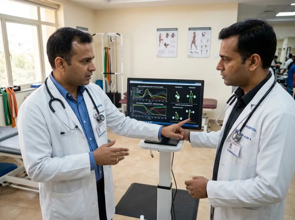
A vector coding study compares elite and sub-elite weightlifters during maximal snatch attempts, revealing that performance differences stem from phase-dependent knee-ankle coordination during transition and turnover phases rather than overall trajectory patterns.
Today

A pilot feasibility study demonstrates that a newly developed mobile app for bolus insulin dose adjustment significantly improves dosing confidence, treatment adherence, and hypoglycemia prevention for patients with type 1 diabetes while reducing provider workload in resource-limited clinical settings.
Today
