

Loading, please wait...
Loading, please wait...

Mechanical thrombectomy represents a cornerstone intervention for acute ischemic stroke resulting from emergent large vessel occlusions. However, post-procedural hemorrhagic transformation remains a feared complication that profoundly threatens functional recovery. Clinicians often encounter persistent hyperdense parenchymal areas on immediate post-intervention cranial imaging. These hyperattenuations typically represent either genuine petechial hemorrhage or benign iodine extravasation caused by disrupted blood-brain barrier permeability. Furthermore, conventional single-energy non-contrast scans cannot reliably distinguish between these two phenomena because both substances demonstrate high radiographic attenuation. Consequently, clinicians frequently face severe therapeutic hesitation when deciding on immediate antiplatelet initiation or intensive hemodynamic targets. To address this persistent clinical challenge, dual-energy computed tomography has emerged as a revolutionary modality capable of spectral tissue characterization. By exploiting the distinct photon absorption properties of materials across diverse energy levels, this advanced technique enables prompt, reliable post-recanalization evaluation.
Endovascular recanalization restores critical cerebral perfusion, yet it simultaneously exposes fragile microvasculature to renewed hydrostatic pressure. As a result, blood-brain barrier disruption frequently permits the transmigration of radiographic contrast into infarcted tissues. When patients undergo standard cranial computed tomography immediately following thrombectomy, radiologists frequently detect dense parenchymal staining. Unfortunately, standard Hounsfield unit measurements cannot reliably separate hyperdense contrast medium from hyperacute petechial bleeding. This diagnostic ambiguity creates a considerable dilemma for neurocritical care teams. If clinicians assume that contrast staining is an evolving hematoma, they may inappropriately withhold necessary antiplatelet or anticoagulant agents. Conversely, mistaking genuine intracranial hemorrhage for benign contrast pooling can lead to catastrophic delays in blood pressure lowering and secondary neuroprotection. Therefore, physicians urgently require an immediate, definitive diagnostic modality directly following endovascular procedures. Establishing certainty within the first postoperative hour significantly optimizes neurointensive workflows and preserves vulnerable brain tissue.
Spectral imaging technology effectively resolves the longstanding diagnostic conundrum of contrast extravasation versus acute hematoma. Specifically, dual-energy computed tomography simultaneously acquires two distinct radiographic datasets using two separate energy spectra, typically 80 to 100 kilovolts and 140 kilovolts. Because iodine and hemoglobin exhibit radically different atomic numbers, their photoelectric absorption behaviors diverge markedly at differing photon energies. Advanced post-processing algorithms utilize this physical principle through three-material decomposition models. Consequently, the scanner generates virtual non-contrast reconstructions and quantitative iodine maps from a single examination. The virtual non-contrast algorithm mathematically subtracts iodine attenuation, unmasking underlying parenchymal hemorrhage with exquisite clarity. Meanwhile, the iodine overlay map highlights isolated contrast pooling without structural parenchymal disruption. Therefore, neurointerventional teams no longer need to wait twenty-four hours for contrast clearance to determine actual tissue status. Furthermore, this spectral decomposition avoids additional radiation exposure compared to repetitive serial conventional examinations.
A comprehensive clinical study evaluated the real-world diagnostic accuracy of immediate postoperative dual-energy imaging in acute stroke patients. Specifically, investigators retrospectively analyzed 118 consecutive stroke patients who underwent dual-energy computed tomography immediately following mechanical thrombectomy. The clinical team conducted follow-up cranial imaging on the subsequent day using either conventional tomography or magnetic resonance imaging to verify findings. Among the 118 evaluated patients, immediate spectral scans demonstrated suspected bleeding in 39 individuals. Subsequent next-day imaging confirmed actual hemorrhagic transformation in 37 of these 39 patients. Notably, only two cases proved to be false positives, where hyperdensities resolved without hemorrhagic evolution. Additionally, the imaging protocol missed only a single hemorrhagic event, representing an isolated false-negative outcome. Overall, the technique demonstrated an exceptional diagnostic sensitivity of 97.4% and an outstanding specificity of 97.5%. Consequently, these compelling metrics confirm that immediate spectral scanning provides nearly definitive diagnostic assurance for neurovascular teams directly following revascularization.
The exceptional diagnostic precision of immediate spectral scanning directly influences hemodynamic and pharmacologic protocols in the neuro-intensive care unit. For instance, clinical guidelines typically advise stringent blood pressure reduction to prevent hematoma expansion when acute bleeding occurs. However, overly aggressive blood pressure lowering in patients without hemorrhage might compromise cerebral perfusion in marginal collateral territories. Because spectral imaging rapidly rules out hemorrhage, clinicians can safely maintain permissive or individualized blood pressure targets to sustain viable penumbral tissue. Furthermore, post-thrombectomy protocols often demand the rapid deployment of antiplatelet therapies, particularly after intracranial stenting or carotid angioplasty. Withholding antiplatelets due to ambiguous contrast staining risks acute stent thrombosis and catastrophic vessel re-occlusion. Therefore, confirming the absence of true extravasated blood allows clinicians to start indicated antiplatelet regimens with greater therapeutic confidence. In contrast, early identification of actual hemorrhage triggers prompt antiplatelet suspension and strict systolic pressure titration.
Although dual-energy computed tomography provides superior diagnostic performance, neurointerventionists and radiologists must recognize potential technical limitations. In the evaluated study cohort, two false-positive findings occurred, reminding clinicians that post-processing artifacts can occasionally simulate true parenchymal bleeding. For instance, extreme iodine concentrations or localized metal artifacts from metallic clips, coils, or dental hardware can introduce material decomposition errors. Furthermore, motion artifacts caused by restless or agitated stroke patients can distort voxel registration across energy spectra. When algorithm subtraction becomes imperfect, small areas of residual iodine hyperattenuation might incorrectly suggest underlying blood elements. However, clinicians can minimize these errors by evaluating both the virtual non-contrast images and the corresponding iodine maps side-by-side. Additionally, quantitative measurement of iodine concentration provides objective validation, distinguishing dense extravasation from genuine hematoma. Thus, combining disciplined qualitative assessment with quantitative attenuation thresholds ensures that clinical teams avoid diagnostic misinterpretations and unwarranted management changes.
Implementing immediate spectral scanning into institutional acute stroke algorithms enhances overall patient outcomes and hospital efficiency. When medical centers integrate this diagnostic protocol into their comprehensive stroke services, patient transport pathways become streamlined and safe. In practice, patients transition directly from the catheterization laboratory to the adjacent spectral scanner before transferring to the neurocritical care unit. Consequently, neurointensivists receive decisive structural and vascular data within minutes of procedural completion. This operational efficiency eliminates the clinical anxiety associated with conventional watchful-waiting strategies that delay medical management for 24 hours. Moreover, early definitive classification enables nursing teams to tailor specialized neuromonitoring protocols based on objective bleeding risks. Looking forward, the wider availability of multi-energy systems in tertiary centers will expand access to this essential diagnostic capability. As clinical evidence continues to accumulate, immediate dual-energy computed tomography will likely solidify its role as an indispensable standard of care in neuroendovascular intervention.
Dual-energy imaging scans tissues simultaneously using high and low kilovoltage spectra. Because iodine has a high atomic number, its radiographic attenuation changes markedly between energies compared to biological blood. Three-material decomposition software calculates these specific energy differences, mathematically subtracting iodine to produce virtual non-contrast images that cleanly unmask true hemorrhage.
Clinicians should ideally perform spectral imaging immediately after mechanical thrombectomy before transferring the patient to the intensive care unit. This immediate assessment distinguishes benign contrast extravasation from acute petechial hemorrhage within minutes. Consequently, critical care teams can promptly optimize antithrombotic therapies and establish safe hemodynamic parameters without unnecessary delays.
Immediate detection of intracranial bleeding prompts intensive blood pressure reduction, typically maintaining systolic levels below 140 mmHg to curb hematoma expansion. Conversely, if spectral imaging confirms benign contrast staining without true hemorrhage, clinicians can safely avoid overly aggressive blood pressure drops, thereby preserving collateral perfusion to vulnerable peri-infarct brain tissue.
Disclaimer: This content is for informational and educational purposes only. It is not intended to substitute professional medical advice, diagnosis, or treatment. Medical knowledge and clinical guidelines constantly change. Clinicians should use their clinical judgment and cross-reference multiple validated references before making therapeutic decisions. The authors and publishers assume no liability for any injury or damage resulting from the use of information contained in this material. Refer to the latest local and national guidelines for clinical practice.
References

Read summarized clinical updates, watch expert medical content, and earn CME certifications right from your smartphone.
Open your smartphone camera to scan and install.





A retrospective cohort study demonstrates that immediate dual-energy computed tomography achieves 97.4% sensitivity and 97.5% specificity in distinguishing intracerebral hemorrhage from contrast extravasation after mechanical thrombectomy, optimizing post-stroke blood pressure control and secondary prevention.
Today

A breakthrough ultrathin multimodal sensor decouples physical hemodynamic monitoring from disposable electrochemical calibration, achieving long-term wearable glucose prediction with 81.36% accuracy and a 9.12% MARD. This offers a robust, drift-free solution for outpatient metabolic surveillance.
Today

A clinical review of menopause and neurology highlights the neurobiological impacts of midlife hormonal shifts on cognition, epilepsy, migraine, multiple sclerosis, and stroke, emphasizing safe hormone therapy prescribing.
Today

Co-administering antiseizure medications and DOACs frequently causes pharmacokinetic interactions, subtherapeutic drug levels, and thromboembolic failures. This review highlights enzyme induction risks, unpredictable outcomes with levetiracetam, and the vital role of therapeutic drug monitoring.
Today
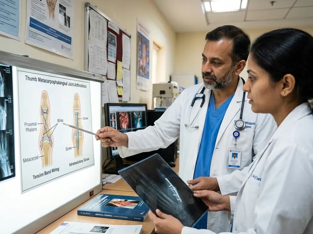
A comparative review of tension band wiring and intramedullary screw fixation in thumb metacarpophalangeal arthrodesis, evaluating union rates, hardware complications, and clinical outcomes.
Today

Following regulatory scrutiny from FSSAI regarding promotional representations and lean muscle preservation claims, Dr. Reddy's Laboratories and Nestlé Health Science are currently reviewing the branding and marketing strategy for their joint nutritional supplement Celevida GLP+.
Today
