.webp&w=384&q=75)

Loading, please wait...
Loading, please wait...
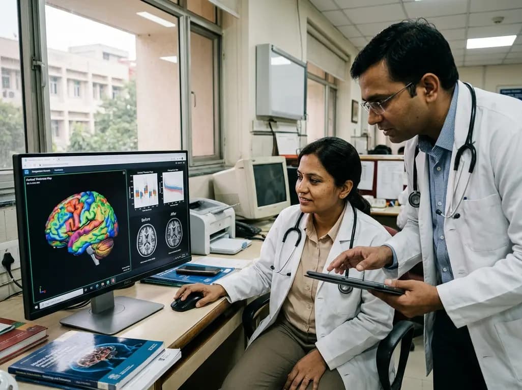
Neuroimaging biomarkers have revolutionized the diagnostic and prognostic landscapes across neurological disciplines. Specifically, precise cortical thickness estimates derived from magnetic resonance imaging (MRI) serve as vital metrics for tracking neurodegenerative changes, cortical thinning, and disease trajectory in conditions such as multiple sclerosis and Alzheimer's disease. However, standardizing these morphometric measurements across diverse clinical settings remains challenging because variations in sequence parameters introduce substantial measurement artifacts. Addressing these discrepancies is essential for translating advanced computational neuroimaging into routine clinical workflows.
Quantitative MRI morphometry heavily relies on consistent image contrast to segment neuroanatomical structures accurately. In multi-center clinical trials and everyday clinical radiology, technical parameters such as repetition time (TR) and inversion time (TI) frequently fluctuate across different scanner vendors and acquisition protocols. Consequently, these technical discrepancies alter tissue contrast ratios at the gray-white matter interface, introducing artificial variances into volumetric calculations. Therefore, longitudinal patient tracking often suffers because true biological changes become confounded by scanner-induced variability.
Traditional segmentation pipelines such as FreeSurfer frequently demonstrate vulnerability to these subtle parameter shifts. Although these tools offer established analytical frameworks, their computational dependency on specific intensity distributions limits their generalizability. Furthermore, conventional deep learning models trained on uniform datasets frequently overfit to homogeneous contrast profiles, failing when deployed across heterogeneous hospital networks. As a result, clinicians and clinical researchers encounter difficulties when comparing scans acquired across different imaging centers or over extended observation windows. Developing automated tools that withstand sequence shifts without losing pathological sensitivity represents a crucial milestone in modern neuroimaging.
To overcome contrast-induced instability, neuroimaging researchers fine-tuned DL+DiReCT, an advanced deep-learning framework combining neural segmentation with diffeomorphic registration. Specifically, the team trained the underlying convolutional architecture using simulated Magnetization Prepared Rapid Gradient Echo (MPRAGE) scans derived from comprehensive quantitative relaxation maps. By exposing the network to a broad spectrum of simulated inversion and repetition times during training, the model learned true morphological geometries rather than superficial intensity boundaries. Consequently, this domain randomization strategy effectively decoupled tissue classification from sequence-dependent contrast variations.
The fine-tuning process dramatically enhanced parameter resilience across rigorous experimental evaluations. For instance, the Pearson correlation coefficient reflecting contrast sensitivity decreased markedly, demonstrating that the fine-tuned architecture operates independently of sequence fluctuations. Moreover, this improved contrast invariance did not compromise processing efficiency or spatial resolution. DL+DiReCT preserved the fine anatomical details of cortical sulci and gyri, enabling rapid and reliable computation. Consequently, this methodological advancement offers clinicians an automated pipeline capable of delivering accurate cortical thickness estimates despite disparate scanning protocols.
Validating deep learning models against ground truth atrophy represents a fundamental requirement prior to clinical adoption. To evaluate performance rigorously, investigators tested the fine-tuned framework against a dedicated synthetic atrophy dataset. In this rigorous evaluation, the fine-tuned DL+DiReCT accurately replicated progressive atrophy trajectories while exhibiting minimal underestimation of true cortical loss. In contrast, standard analytical pipelines displayed notable susceptibility to sequence-induced confounding, frequently miscalculating subtle tissue reductions.
Specifically, the fine-tuned network clearly outperformed both FreeSurfer and SynthSeg in tracking synthetic gray matter volume loss across simulated parameter variations. While SynthSeg provides contrast-agnostic segmentation, DL+DiReCT demonstrated superior sensitivity to delicate boundary shifts, capturing sub-millimeter decrements without generating segmentation noise. Furthermore, the model avoided artificial smoothing, which commonly obscures focal cortical lesions or regional thinning. Therefore, these benchmark results confirmed that eliminating contrast dependence does not diminish the model's capacity to detect true biological degeneration.
The ultimate test of any neuroimaging biomarker lies in its real-world clinical application across patient cohorts. Accordingly, investigators applied the fine-tuned model to an authentic clinical dataset comprising patients diagnosed with relapsing-remitting multiple sclerosis (RRMS). In this clinical cohort, the model demonstrated a substantial reduction in contrast sensitivity, mirroring the robust results observed in synthetic evaluations. Importantly, the framework delivered reliable cortical thickness measures across diverse scanner field strengths and acquisition variations.
Furthermore, the model maintained consistent performance after statistically controlling for critical clinical covariates, including patient age, sex, scanner field strength, and Expanded Disability Status Scale (EDSS) scores. Consequently, the measured cortical thinning reflected authentic neurodegenerative progression rather than technical noise or demographic confounding. Because cortical gray matter atrophy strongly correlates with long-term disability accumulation in RRMS, having a dependable biomarker enables clinicians to assess therapeutic efficacy more objectively. Thus, the fine-tuned pipeline provides actionable prognostic insights that can refine patient management strategies.
Multi-center clinical trials and longitudinal observational studies frequently face significant logistical hurdles regarding MRI protocol harmonization. When trials span multiple years and hospital sites, hardware upgrades and software updates inevitably introduce sequence shifts that jeopardize longitudinal biomarker comparisons. Because the fine-tuned DL+DiReCT tool exhibits intrinsic contrast invariance, it minimizes the necessity for complex retrospective statistical harmonization techniques. Therefore, retrospective and prospective clinical studies can pool neuroimaging data more seamlessly.
Additionally, this technological advancement reduces the sample sizes required to achieve statistical power in clinical trials evaluating neuroprotective therapies. When measurement noise from sequence discrepancies is eliminated, subtle reductions in atrophy rates become readily detectable. Moreover, the computational efficiency of deep learning allows rapid batch processing of large imaging databases, accelerating discovery workflows. Consequently, clinical researchers can evaluate experimental disease-modifying therapies with greater confidence, knowing that structural endpoints reflect true therapeutic responses rather than technical variability across participating medical centers.
Integrating robust AI models into routine Picture Archiving and Communication Systems (PACS) represents the next frontier in diagnostic neuroradiology. As deep learning algorithms mature, automating cortical thickness quantification will empower clinicians to detect early neurodegeneration before irreversible neurological deficits manifest. Furthermore, combining cortical thickness metrics with advanced fluid biomarkers and clinical assessments will enrich precision medicine in neurology.
However, continuous external validation across broader ethnodemographic populations and diverse scanner manufacturers remains essential. Clinicians and radiologists must collaborate to ensure these algorithmic tools integrate smoothly into existing radiological reporting workflows. By establishing contrast-invariant pipelines, the medical imaging community moves closer to standardized, objective neurodegenerative biomarkers that improve diagnostic confidence and patient outcomes globally.
Variations in sequence parameters like repetition time and inversion time alter tissue contrast at gray-white matter interfaces. Consequently, standard automated segmentation tools misinterpret these intensity changes as actual biological differences, generating artificial measurement errors that confound longitudinal tracking and multi-center study data.
The model achieves contrast invariance through fine-tuning on simulated MPRAGE scans derived from quantitative relaxation maps. By training on a wide variety of simulated sequence parameters, the neural network learns underlying neuroanatomical geometry rather than relying solely on specific image contrast profiles.
In multiple sclerosis, cortical gray matter atrophy strongly correlates with permanent physical disability and cognitive decline. Accurately measuring cortical thinning across longitudinal scans enables clinicians to evaluate disease progression objectively and monitor therapeutic responses to disease-modifying therapies without scanner-induced confounding.
Disclaimer: This content is for informational and educational purposes only and is intended for healthcare professionals. It should not be used as a substitute for professional medical advice, diagnosis, or treatment. Refer to the latest local and national guidelines for clinical practice.
References

Read summarized clinical updates, watch expert medical content, and earn CME certifications right from your smartphone.
Open your smartphone camera to scan and install.





Fine-tuning deep-learning segmentation models on simulated MPRAGE images reduces contrast sensitivity and boosts the accuracy of cortical thickness measurements across variable MRI sequence parameters, advancing neurodegenerative biomarker precision in conditions like multiple sclerosis.
last month

Top-down CSF peptidomics uncovers novel endogenous peptide signatures and oxidative modifications differentiating multiple sclerosis from NMOSD, highlighting secretogranin-5 loss as a key biomarker of disease progression.
Today
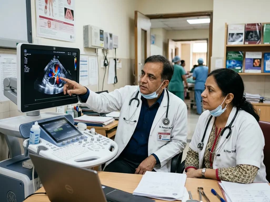
Fetal foramen ovale restriction typically emerges in the third trimester with right heart enlargement. Evidence demonstrates it does not independently mandate cesarean delivery, and over 93% of postnatal cardiac findings resolve spontaneously within one year, supporting conservative obstetric management.
Today

The latest WHO Global Health Estimates highlight India's life expectancy reaching 71 years alongside a healthy lifespan of 60.6 years. While longevity rebounds post-pandemic, non-communicable conditions now account for three-quarters of top mortalities, underscoring urgent clinical shifts in routine primary care.
Today

New biomechanical research reveals that foot-strike angle significantly influences knee energy absorption during cutting tasks in female soccer players. Understanding these kinematic patterns offers clinicians valuable pathways for targeted ACL injury prevention and athletic retraining.
Today

A comprehensive review analyzes periconceptional GLP-1 receptor agonist exposure, revealing reassuring data on congenital anomalies and adverse perinatal outcomes while emphasizing the need for cautious patient counseling.
Today
