.webp&w=384&q=75)

Loading, please wait...
Loading, please wait...
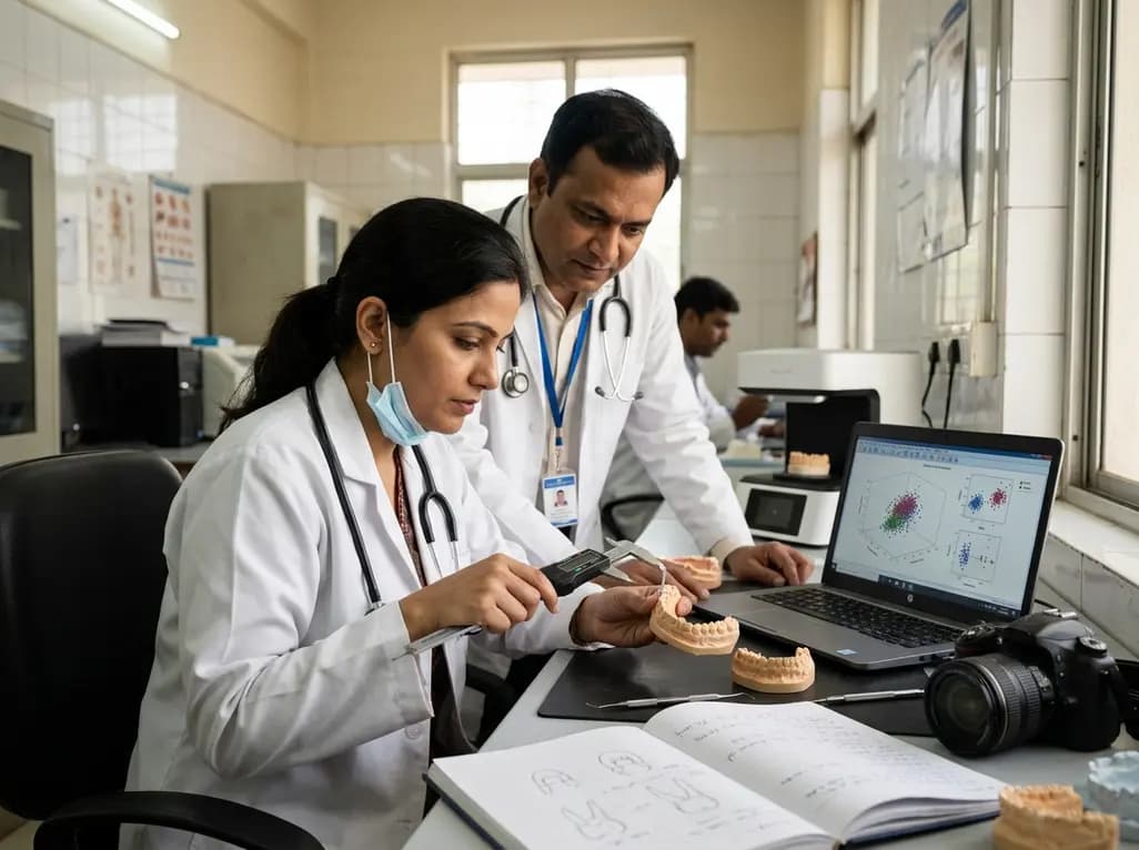
Establishing biological identity from skeletal and dental remains represents a fundamental pillar of forensic science and clinical anthropology. While pelvic and cranial morphology traditional serve as primary indicators for sex determination in adult remains, forensic specialists often face substantial hurdles when evaluating adolescent individuals. During adolescent growth phases, traditional skeletal features may remain incompletely developed or morphologically ambiguous. Digital odontometric analysis has emerged as a reliable, non-invasive alternative that addresses these diagnostic challenges by evaluating human dentition, which exhibits high resistance to postmortem degradation, thermal stress, and environmental trauma.
Human teeth complete crown morphogenesis relatively early in developmental life, preserving stable dimensional traits that reflect genetic sex dimorphism. Recent technological advances in three-dimensional surface scanning and virtual dental morphometry have modernized forensic dentistry. Rather than relying on traditional manual calipers that risk damaging delicate physical specimens, digital odontometric analysis utilizes virtual dental models to capture micrometric dimensional variations across various dental arches. This digital paradigm enhances intra-observer reliability, facilitates multi-center data sharing, and allows complex spatial measurements that were previously unachievable through conventional clinical techniques. Implementing precise digital workflows significantly improves identification accuracy in both forensic investigations and specialized pediatric dental contexts.
To evaluate the scientific precision of digital odontometric analysis, researchers digitized a cohort of 150 high-quality dental casts obtained from Egyptian adolescents aged between 12 and 19 years. Utilizing high-resolution three-dimensional optical scanners, investigators created detailed virtual representations of upper and lower arch relationships. Specialized orthomorphic tracing software was subsequently employed to record linear and transverse dental parameters with high reproducibility. Specific measurements included mesiodistal and buccolingual crown dimensions of the permanent first molars, alongside mesiodistal widths of the maxillary permanent canines.
Beyond individual tooth dimensions, the study systematically documented comprehensive dental arch parameters, including intercanine, interpremolar, and intermolar widths across the maxillary and mandibular dental arches. Digitizing these anatomical structures eliminated operator bias associated with physical placement of manual measurement tools. Advanced statistical comparisons were performed to detect sexual dimorphism across all collected variables. Discriminant function analysis was then applied to construct predictive mathematical models specifically calibrated for adolescent populations. These computational models process multi-parametric dental measurements simultaneously, providing objective probabilistic estimations of individual sex. The methodology highlights how high-resolution digital scanning combined with precise software metrics creates standardized, reproducible forensic protocols.
Statistical evaluation revealed significant sexual dimorphism across all measured dental parameters within the Egyptian adolescent study population. Male subjects consistently demonstrated significantly higher mean values compared with female subjects across mesiodistal crown diameters, buccolingual widths, and overall arch metrics. The permanent maxillary canine displayed pronounced dimorphic characteristics, reinforcing its established status as one of the most sexually dimorphic teeth in the human dentition. Similarly, the permanent first molars exhibited notable dimensional differences, particularly in their buccolingual measurements, which reflect differential enamel thickness and underlying dentinal growth pattern differences between biological sexes.
Transverse arch dimensions, including intercanine and intermolar distances, further contributed to biological differentiation. Male dental arches reflected broader transverse alignment, consistent with generalized craniofacial growth trends observed during pubertal development. By combining these individual dental measurements with arch width metrics through multivariate discriminant function analysis, researchers achieved impressive diagnostic accuracy for sex estimation. The constructed mathematical models achieved high predictive sensitivity and specificity, confirming that digital odontometric analysis yields sufficient discriminatory power to serve as a primary or auxiliary biological profiler when assessing adolescent dental remains in forensic contexts.
The clinical and medico-legal implications of standardized digital odontometric analysis extend far beyond historical population research. In disaster victim identification scenarios, mass casualty events, and medico-legal investigations where skeletal elements are fragmented or incomplete, dentition often provides the sole remaining source of intact biological data. Establishing population-specific normative databases is vital for forensic accuracy, as odontometric dimorphism varies across different ethnic and geographic ancestries. Validating these predictive models in an Egyptian adolescent population offers vital local standard reference metrics, mitigating errors caused by applying mismatched international standard tables.
Furthermore, integrating digital three-dimensional models into routine pediatric dentistry, orthodontics, and maxillofacial surgery creates opportunities for dual-use clinical data. Virtual arch models captured during routine orthodontic diagnosis can serve as verified antemortem records. In clinical practice, understanding sexual dimorphism in arch dimensions and tooth proportions assists orthodontists in planning customized arch expansion, space maintenance, and aesthetic reconstructive procedures tailored to gender-specific developmental trajectories. The seamless integration of digital scanning technologies thereby bridges the gap between everyday clinical dental practice and formal forensic identification protocols.
As digital health technology rapidly expands globally, integrating digital odontometric analysis into forensic dentistry workflows represents a major step forward. Modern artificial intelligence and machine learning algorithms can be trained on vast datasets of digitized dental arches to automate feature extraction and sex estimation, further reducing analytical processing times and removing inter-observer variability. Combining automated digital odontometry with advanced imaging modalities, such as cone-beam computed tomography, provides comprehensive three-dimensional visualization of crown morphology, root structures, and surrounding alveolar bone anatomy.
For medical educators, forensic pathologits, and dental practitioners in India, adopting standardized digital odontometric frameworks offers strong potential for enhancing national forensic infrastructure. Developing population-specific digital odontometric databases tailored to diverse Indian ethnicities will strengthen indigenous medico-legal capabilities. Promoting interdisciplinary collaboration between pediatric dentists, oral radiologists, and forensic anthropologists ensures that modern digital tools are effectively utilized for patient care, anatomical research, and humanitarian identification efforts nationwide.
Digital odontometric analysis involves using three-dimensional optical scanners and specialized software to measure dental crown dimensions and arch widths on virtual models. It assists in sex estimation by detecting biological differences in tooth size and arch geometry between males and females, offering an objective, non-invasive forensic tool when skeletal indicators are unavailable.
Permanent canines and first molars exhibit the highest degree of sexual dimorphism in human dentition. Canines show significant differences due to evolutionary and hormonal influences on tooth development, while first molars erupt early and possess durable enamel thickness, making both tooth types exceptionally reliable for forensic sex determination.
Digital scanning eliminates physical measurement errors caused by manual calipers, prevents accidental damage to fragile specimens, and enables precise measurement of complex three-dimensional arch curves. Virtual models can be archived permanently, analyzed remotely by multiple experts, and integrated easily into automated mathematical software for rapid discriminant function analysis.
Disclaimer: This content is for informational and educational purposes only and does not constitute professional medical, dental, or legal advice. Refer to the latest local and national guidelines for clinical and forensic practice.
References
Hadwa SM et al. Digital odontometric analysis for sex estimation in Egyptian adolescents. BMC Oral Health. 2026 Aug 08. doi: undefined. PMID: 42568081.

Read summarized clinical updates, watch expert medical content, and earn CME certifications right from your smartphone.
Open your smartphone camera to scan and install.





A study validates digital odontometric analysis for sex estimation in Egyptian adolescents using 3D scanning and discriminant function analysis. Measurements of permanent molars, canines, and arch widths demonstrated significant sexual dimorphism, offering a non-invasive tool for forensic identification.
last month
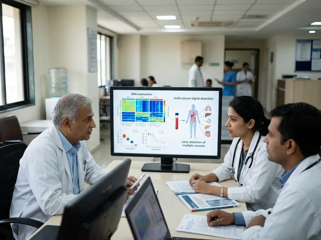
FDA staff reviewers have raised no major safety or analytical concerns regarding Grail's Galleri blood test ahead of an advisory panel meeting. This milestone advances liquid biopsy technologies designed to identify tumor methylation signals across dozens of malignancies before clinical symptoms emerge.
Today

Meghalaya recorded a historic 55% reduction in maternal deaths, dropping from 244 to 107 over five years. Through the MOTHER app, dedicated transit homes, and expanded infrastructure like the 150-bed Tura Maternity Hospital with its first public NICU, the state demonstrates an effective public health model.
Today

Explore modern perioperative management protocols for clinical xenotransplantation. Learn how costimulation blockade, anti-inflammatory perfusion, and coagulopathy rescue overcome interspecies hurdles.
Today
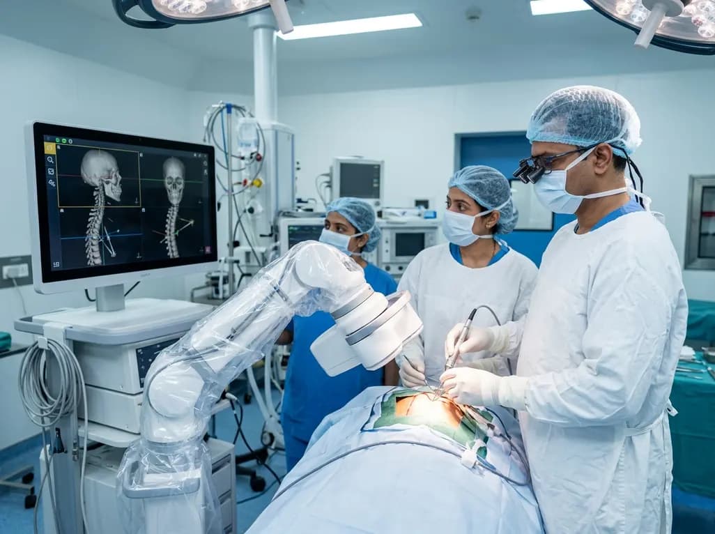
North India's first Centre of Excellence for Brain and Spine at Amrita Hospital Faridabad integrates Brainlab's Loop-X mobile robot. This landmark facility combines cone-beam computed tomography with surgical navigation to elevate precision, optimize operative workflows, and enhance specialized clinician training.
Today

At Homa CME 2026 at Hyderabad's T-Hub, healthcare leaders emphasized moving beyond routine metrics through digital calculators and precision stratification. Discover why characterizing individual insulin secretion, resistance patterns, and vascular risks transforms chronic metabolic care in India.
Today
