.webp&w=384&q=75)

Loading, please wait...
Loading, please wait...

Intracranial meningiomas represent the most frequently encountered primary central nervous system tumors in adult clinical neurosurgery. Over recent decades, widespread availability and rapid adoption of high-resolution magnetic resonance imaging have significantly elevated incidental detection rates. Consequently, clinicians routinely identify asymptomatic or minimally symptomatic tumors in diverse patient populations across outpatient clinics. Despite this rising diagnostic frequency, long-term clinical data reveal unexpected shifts in therapeutic management paradigms. Recent academic research demonstrates that intracranial meningioma surgical incidence has experienced a distinct age-adjusted decline over a nineteen-year period. This notable phenomenon highlights a major transition in neurosurgical philosophy from proactive prophylactic resection toward watchful waiting and individualized risk stratification. Surgeons now weigh surgical risks against true natural disease progression before offering invasive intervention.
A retrospective analysis spanning nearly two decades across more than two thousand consecutive adult patients offers comprehensive insights into contemporary surgical management patterns. Although absolute annual surgical caseloads remained relatively stable over time, standardizing these metrics to standardized population figures demonstrates a consistent downward trajectory in surgical rates across specific age cohorts. This age-adjusted decrease occurred despite population aging and persistent growth in overall neuroimaging availability throughout health system networks. Consequently, researchers confirmed that surgical resection rates operate independently of background tumor detection frequency. Furthermore, Observed-to-Expected ratio analysis reinforced this finding, demonstrating a pronounced reduction in intervention rates among middle-aged and older individuals. This shift highlights a modern consensus among neurosurgical multidisciplinary teams. Today, surgical teams selectively offer resection to patients who face clear clinical necessity rather than operating on incidentally identified, indolent mass lesions. Ultimately, these findings reassure clinicians that increased imaging sensitivity does not necessitate excessive surgical intervention. Instead, neurosurgeons successfully apply evidence-based selection criteria to prevent unnecessary craniotomies while preserving overall patient safety, neurological status, and functional quality of life.
Demographic analysis over the nineteen-year study period reveals a marked increase in the median age of patients undergoing surgical resection. Specifically, the median age of surgical candidates increased by nearly three months each calendar year. This demographic shift reflects overall population aging and demonstrates that advanced age alone no longer acts as an absolute contraindication to complex skull base or convexity surgery. Furthermore, modern neuroanesthesia protocols and improved perioperative critical care allow safe surgical intervention in appropriately selected elderly individuals. However, operating on older adults requires comprehensive preoperative risk assessment to evaluate systemic comorbidities, fragile cerebral vasculature, and baseline functional performance. Modern clinical practice balances chronological age against physiological status when determining surgical eligibility. While older patients present unique physiological challenges, surgical intervention remains highly effective when tailored to specific functional goals. Consequently, neurosurgeons reserve craniotomy for older adults who experience significant mass effect, progressive focal neurological deficits, or refractory intracranial hypertension. By contrast, asymptomatic elderly patients with incidental meningiomas are managed conservatively with serial neuroimaging. This prudent approach protects fragile individuals from surgical complications while preserving baseline independence over time.
Surgical indications for intracranial meningiomas have undergone profound changes over recent decades. Historically, neurosurgeons frequently performed prophylactic resection on small, asymptomatic lesions to prevent potential future growth or neurological deterioration. However, contemporary longitudinal data demonstrate a statistically significant reduction in prophylactic surgical procedures. Today, symptom relief represents the single most common indication for surgical intervention, accounting for nearly two-thirds of all resections. Surgeons now prioritize operation for patients presenting with documented neuroimaging growth, prominent mass effect, or significant neurological dysfunction. Conversely, incidental discovery without accompanying clinical symptoms rarely prompts immediate operative resection. Furthermore, clinical evidence confirms that many small meningiomas exhibit indolent growth rates or complete growth arrest over extended observation periods. Therefore, modern guidelines emphasize conservative active surveillance with annual magnetic resonance imaging for non-symptomatic tumors. This selective strategy prevents unnecessary surgical morbidity while ensuring timely intervention if objective radiological progression or clinical deterioration occurs. Consequently, the threshold for surgical resection has become considerably more stringent across academic medical centers worldwide, benefiting overall patient management.
Understanding presenting symptoms is essential for establishing appropriate management strategies for patients diagnosed with intracranial meningiomas. Clinical registry data indicate that new-onset seizures and visual disturbances constitute the most frequent presenting complaints leading to neurosurgical consultation. Additionally, patients may experience localized headaches, focal motor deficits, cranial nerve palsies, or cognitive alterations depending on tumor location and surrounding cerebral edema. When patients present with severe, debilitating symptoms, surgical resection offers immediate relief of mass effect and marked clinical improvement. Conversely, when patients present with incidental findings during evaluation for unrelated complaints, conservative management becomes the preferred primary approach. Active surveillance protocols involve sequential magnetic resonance imaging to track volumetric changes over time. If follow-up imaging reveals stable tumor dimensions without surrounding vasogenic edema, neurosurgeons safely continue conservative observation. Furthermore, this monitoring approach enables clinicians to identify true biological aggressiveness before committing patients to surgical risk. Ultimately, detailed symptom mapping and routine imaging surveillance allow neurosurgeons to identify the optimal therapeutic window for operative intervention when necessary, preserving cerebral function.
The rapid evolution of diagnostic neuroimaging has revolutionized the clinical approach to intracranial meningiomas. Advanced MRI techniques, including perfusion imaging, diffusion tensor imaging, and MR spectroscopy, provide critical information regarding tumor vascularity, cellularity, and adjacent white matter tract displacement. Furthermore, functional MRI helps neurosurgeons map eloquent cortical areas relative to the tumor capsule prior to surgical planning. These sophisticated technological advances allow clinical teams to differentiate indolent lesions from aggressive biological subtypes with greater accuracy. Consequently, surgeons can confidently recommend conservative observation for low-risk, radiologically stable lesions without compromising long-term patient outcomes. Moreover, advanced neuroimaging facilitates precise surgical planning when resection is required, improving complete resection rates while minimizing neurological deficits. The integration of volumetric image analysis also enables precise quantitative tracking during active surveillance protocols. By replacing subjective diameter measurements with accurate three-dimensional volumetric assessments, clinicians detect true tumor growth earlier. Therefore, neuroimaging advances serve a dual purpose: avoiding unneeded operations in stable patients and optimizing operative precision for those needing surgical resection.
The observed decline in age-adjusted surgical incidence reflects a major paradigm shift toward individualized, risk-stratified patient management. Modern neurosurgical decision-making synthesizes clinical presentation, patient preferences, comorbidities, tumor location, and radiological growth kinetics into a cohesive therapeutic strategy. Rather than applying uniform surgical criteria, multidisciplinary neuro-oncology teams carefully evaluate each patient's personalized risk-benefit profile. Furthermore, non-surgical alternatives such as stereotactic radiosurgery and fractionated radiation therapy offer highly effective options for select patients. Radiotherapy provides durable local tumor control for deep-seated skull base lesions or medically frail individuals who face elevated surgical risks. Moreover, ongoing molecular and genomic research promises to refine risk stratification further by identifying specific genetic alterations associated with aggressive tumor behavior. As molecular profiling becomes routinely available in neuro-oncology practice, neurosurgeons will increasingly tailor observation, surgery, or targeted adjuvant therapies to specific tumor biology. Consequently, modern neurosurgical practice continues to move away from aggressive prophylactic surgery toward highly selective, patient-centered care that maximizes long-term functional independence, safety, and quality of life.
The decline in age-adjusted surgical incidence reflects a clinical paradigm shift toward selective, risk-stratified management. Rather than operating prophylactically on incidentally detected tumors, neurosurgeons now reserve surgery for cases demonstrating significant clinical symptoms, documented radiological growth, prominent mass effect, or focal neurological deficits, prioritizing watchful waiting for indolent lesions.
Symptom relief represents the single most common surgical indication for intracranial meningioma. Resection is primarily recommended for patients presenting with tumor-related seizures, visual impairment, focal neurological deficits, or substantial mass effect, as well as those demonstrating unambiguous radiological growth during active neuroimaging surveillance protocols.
Advanced age alone is no longer an absolute contraindication to meningioma surgery. However, modern neurosurgical decision-making balances chronological age against physiological status and systemic comorbidities. Asymptomatic elderly patients with stable tumors undergo conservative neuroimaging observation, whereas surgery is selectively offered when progressive symptoms or mass effect threaten independence.
Disclaimer: This content is for informational and educational purposes only and does not constitute medical advice. Refer to the latest local and national guidelines for clinical practice.
References

Read summarized clinical updates, watch expert medical content, and earn CME certifications right from your smartphone.
Open your smartphone camera to scan and install.





A 19-year retrospective analysis reveals declining age-adjusted surgical incidence of intracranial meningiomas despite increased neuroimaging detection. Surgical intervention is increasingly reserved for symptomatic or growing tumors, marking a shift toward selective, risk-stratified patient care.
2 months ago
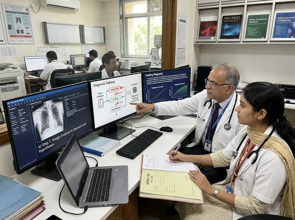
A breakthrough peer-reviewed study demonstrates that a sequential AI diagnosis system can mirror real-world clinical reasoning. By ordering stepwise tests and updating hypotheses dynamically, this multi-agent orchestrator achieved superior diagnostic accuracy while reducing investigative costs.
Today

A prenatal case highlights severe glutaric acidemia type II presenting as bilateral hyperechoic cystic kidneys at 18 weeks. Whole-exome sequencing revealed a novel homozygous ETFDH frameshift variant, demonstrating the power of rapid genetic testing in fetal renal anomalies.
Today

Review how tailored exercise programs benefit frail older patients with heart failure. Explore pathophysiological mechanisms, individualized training prescriptions, and strategies to overcome adherence barriers in clinical practice.
Yesterday

This comparative study evaluates posterior labral tears with and without glenohumeral instability, highlighting distinct demographic patterns, MRI underestimation in stable shoulders, and successful arthroscopic repair outcomes.
Yesterday

A clinical study demonstrates that early intervention with tocilizumab, either as monotherapy or bridged with corticosteroids, achieves significant proptosis reduction and resolves active inflammation in treatment-naive thyroid eye disease.
Yesterday
