.webp&w=384&q=75)

Loading, please wait...
Loading, please wait...

Accurate preoperative identification of rare mesenchymal liver lesions presents a formidable challenge to clinical hepatologists and cytopathologists alike. Specifically, hepatic PEComa represents an uncommon mesenchymal neoplasm displaying distinctive myomelanocytic differentiation. Because clinicians infrequently encounter these lesions in routine hepatology clinics, misdiagnosing them as primary liver carcinomas or secondary metastases occurs frequently. Fine-needle aspiration biopsy, coupled with ancillary immunohistochemistry and molecular analysis, provides an indispensable pathway toward achieving an accurate, non-surgical diagnosis before definitive operative resection.
Perivascular epithelioid cell tumors encompass a distinct group of mesenchymal neoplasms recognized by the World Health Organization. Historically, this tumor family includes angiomyolipoma, clear cell sugar tumor of the lung, and lymphangioleiomyomatosis. In the liver, the epithelioid angiomyolipoma variant constitutes a notable subset that predominantly affects young to middle-aged female patients. Morphologists characterize these lesions by their close association with blood vessel walls and their hybrid phenotype, which combines both melanocytic and smooth muscle attributes.
Furthermore, these neoplasms display a wide spectrum of biological behavior. While the vast majority of these lesions exhibit an indolent clinical course, a small subset demonstrates aggressive biological potential. Consequently, pathologists must rigorously evaluate cellular architecture, nuclear pleomorphism, mitotic activity, and necrosis. In addition, syndromic associations often accompany these neoplasms, particularly tuberous sclerosis complex. However, sporadic cases also occur frequently in patients without syndromic features, as demonstrated in individuals evaluated during routine surveillance following remission from other unrelated malignancies. Therefore, establishing a reliable diagnostic framework remains critical for multidisciplinary oncology teams.
Patients with hepatic PEComa often remain completely asymptomatic during the early stages of tumor growth. Consequently, radiologists and clinicians typically discover these masses incidentally during abdominal imaging or while investigating unexplained liver function test alterations. Dynamic imaging modalities, including contrast-enhanced computed tomography and magnetic resonance imaging, reveal hypervascular features with arterial phase hyperenhancement. Unfortunately, these imaging findings closely mirror the characteristic vascular profile of hepatocellular carcinoma and focal nodular hyperplasia.
Moreover, non-cirrhotic patients who present with arterial enhancement create substantial clinical dilemmas for multidisciplinary teams. Because classic imaging algorithms, such as LI-RADS, rely heavily on underlying cirrhosis, they often fail to categorize mesenchymal neoplasms accurately. Consequently, misdiagnosing these lesions as hepatocellular carcinoma occurs in up to eighteen percent of cases. Additionally, clinicians frequently consider metastatic melanoma, gastrointestinal stromal tumors, and neuroendocrine neoplasms in the differential diagnosis. Therefore, tissue sampling becomes imperative whenever imaging findings deviate from standard diagnostic criteria or occur in atypical clinical demographics. Ultrasound-guided aspiration provides a safe, highly controlled technique for obtaining diagnostic cellular material without requiring invasive exploratory surgery.
Fine-needle aspiration biopsy yields cellular specimens that provide crucial diagnostic clues when evaluated by experienced cytopathologists. Aspirates typically show hypercellular smears featuring discohesive architecture, characterized by singly dispersed neoplastic cells alongside loosely arranged clusters. Cytologists observe both epithelioid and short spindle cells displaying abundant, finely granular to clear eosinophilic cytoplasm. Moreover, tumor cells exhibit central to eccentric nuclei with mild to moderate nuclear pleomorphism. Prominent macronucleoli frequently appear within the nuclei, reflecting intense metabolic activity.
Furthermore, rapid on-site evaluation ensures adequate specimen cellularity and facilitates triage for ancillary diagnostic testing. Cell block preparations remain vital because they allow cytopathologists to evaluate architectural relationships and perform comprehensive immunocytochemical panels. However, pathologists must avoid interpreting moderate nuclear atypia and prominent nucleoli as definitive evidence of high-grade malignancy. Although these cytomorphologic features often raise suspicion for poorly differentiated carcinoma or melanoma, the presence of distinct dual cell populations and clean backgrounds helps guide the interpretation. Thus, careful cytologic examination combined with cell block processing serves as the diagnostic cornerstone for confirming this rare mesenchymal neoplasm.
Immunohistochemical staining remains essential for confirming the diagnosis of hepatic PEComa because cytomorphology alone cannot differentiate it from mimickers. Neoplastic cells characteristically demonstrate dual differentiation, showing strong immunoreactivity for both melanocytic and myogenic markers. Pathologists routinely observe diffuse positivity for Human Melanoma Black 45 and Melan-A alongside smooth muscle actin and caldesmon. Conversely, these tumors consistently lack epithelial markers, including broad-spectrum cytokeratins, and demonstrate negative staining for hepatocyte-specific markers like HepPar-1 and Arginase-1.
Additionally, molecular genetics provides critical diagnostic confirmation and therapeutic insights into the disease process. Pathogenic alterations in the tuberous sclerosis genes, specifically TSC1 and TSC2, drive the underlying pathogenesis of these tumors. Biallelic inactivation of TSC2 disrupts the tuberin-hamartin complex, resulting in constitutive hyperactivation of the mechanistic target of rapamycin signaling pathway. Splice-site and frameshift mutations in TSC2 trigger unabated cellular proliferation and distinct metabolic reprogramming. Furthermore, identifying these molecular alterations carries profound clinical significance. If surgical resection proves impossible due to tumor location or anatomical complexity, clinicians can deploy targeted mTOR inhibitors, such as sirolimus or everolimus, to achieve effective systemic disease control.
Distinguishing hepatic PEComa from morphological mimics requires systematic diagnostic algorithms and multidisciplinary collaboration. Hepatocellular carcinoma represents the most common diagnostic pitfall, especially the clear-cell and pleomorphic variants. However, hepatocellular carcinoma expresses arginase-1, glypican-3, and polyclonal carcinoembryonic antigen while remaining negative for HMB-45. Similarly, metastatic melanoma shares melanocytic marker expression, but it typically shows strong S100 protein reactivity and lacks smooth muscle actin expression. Furthermore, gastrointestinal stromal tumors exhibit CD117 and DOG-1 positivity, which are conspicuously absent in PEComas.
Consequently, once the multidisciplinary team establishes an accurate preoperative diagnosis, complete surgical resection serves as the gold-standard curative treatment. Surgeons typically perform anatomical or non-anatomical partial hepatectomy with clear microscopic margins. Fortunately, complete surgical resection yields excellent long-term prognosis in the majority of patients with localized, non-invasive lesions. Nevertheless, clinicians must establish structured long-term post-resection follow-up protocols because borderline or malignant variants occasionally recur or metastasize. Surveillance incorporates serial cross-sectional abdominal imaging and routine liver function testing over several years. Through meticulous cytological evaluation, targeted immunohistochemistry, and molecular profiling, clinicians can prevent inappropriate oncologic interventions and achieve optimal outcomes.
Fine-needle aspiration biopsy provides a minimally invasive and highly accurate method to sample suspicious liver masses preoperatively. By yielding intact cellular clusters and singly dispersed epithelioid cells, aspiration allows cytopathologists to evaluate characteristic cytomorphologic features. Furthermore, rapid on-site evaluation ensures adequate cell block preparation for necessary ancillary immunohistochemical and molecular studies. Consequently, this technique reliably distinguishes hepatic PEComas from primary carcinomas and metastatic neoplasms, effectively guiding appropriate surgical planning.
Diagnostic confirmation requires demonstrating a dual myomelanocytic immunophenotype on cytologic cell blocks. The neoplastic cells show strong positivity for melanocytic markers, including HMB-45 and Melan-A. Additionally, they express smooth muscle markers like smooth muscle actin and caldesmon. Conversely, the tumor tests negative for epithelial cytokeratins and hepatocellular markers like HepPar-1 and Arginase-1. Therefore, this distinctive immunoprofile conclusively differentiates PEComas from carcinomas and metastatic melanomas.
Molecular testing for TSC2 mutations provides objective biological confirmation of hepatic PEComa, particularly in diagnostically challenging biopsies. Inactivating mutations in TSC2 disrupt the tuberin-hamartin complex, which causes hyperactivation of the downstream mTOR signaling pathway. Furthermore, confirming this pathogenic alteration validates the mesenchymal origin of the lesion and excludes unrelated mimics. Clinically, identifying this genetic driver also provides a viable rationale for utilizing targeted mTOR inhibitors in unresectable or recurrent disease.
Disclaimer: This content is for informational and educational purposes only. It is not intended to substitute for professional medical advice, diagnosis, or treatment. Always seek the advice of your physician or other qualified healthcare provider with any questions you may have regarding a medical condition. Refer to the latest local and national guidelines for clinical practice.
References

Read summarized clinical updates, watch expert medical content, and earn CME certifications right from your smartphone.
Open your smartphone camera to scan and install.





Hepatic PEComas are rare mesenchymal neoplasms with myomelanocytic differentiation. Discover the cytomorphologic features, immunohistochemical profile, and TSC2 molecular alterations that enable accurate preoperative diagnosis using fine-needle aspiration biopsy.
Today

A study shows that the aggregate index of systemic inflammation (AISI) significantly associates with elevated myocardial injury markers, including cardiac troponin T and CK-MB, in patients with coronary heart disease, highlighting its potential utility for cardiovascular risk stratification.
Today
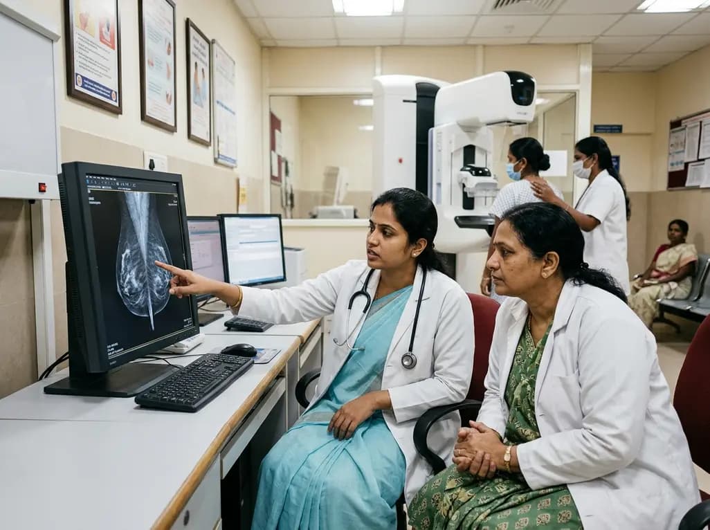
India faces an escalating breast cancer burden marked by late diagnosis and elevated mortality. Shifting from reliance on physical exams to an organized, AI-assisted mammography screening protocol can detect malignancies early, bridge urban-rural disparities, and improve patient survival outcomes across the nation.
Today

A retrospective study reveals that adults with congenital heart disease have significant deficits in peripheral muscle mass and handgrip strength compared to healthy peers. Greater lesion complexity exacerbates muscle loss, demonstrating the need for comprehensive nutritional and muscular assessments beyond standard BMI.
Today

The state government is deploying helicopter emergency medical services across the Mumbai-Pune Expressway, Samruddhi Mahamarg, and Mumbai-Goa Highway. This initiative establishes unified standard operating procedures to enable rapid triage, advanced prehospital resuscitation, and seamless trauma care transfers.
Today

A systematic review of 66 preclinical studies reveals that GLP-1 mono-agonists modulate central reward processing. These findings demonstrate significant reductions in palatable food intake, substance self-administration, and compulsive behaviors, offering novel neurobiological targets for neuropsychiatry.
Today
