.webp&w=384&q=75)

Loading, please wait...
Loading, please wait...

The intersection of tropical parasitic infections and cellular immunodeficiency represents a complex clinical frontier. In particular, managing cutaneous leishmaniasis and HIV coinfection poses extraordinary challenges for clinicians worldwide. When cell-mediated immunity declines due to advanced retroviral disease, protozoan pathogens exhibit heightened virulence, atypical dissemination, and poor response to standard antileishmanial regimens. Recent clinical observations from endemic regions highlight how compromised host defenses dismantle localized containment, allowing localized cutaneous ulcers to transform rapidly into extensive, disseminated polymorphic eruptions. Understanding these dynamics is essential for modern infectious disease practice.
Normally, an immunocompetent host mounts a robust T-helper 1 immune response characterized by interferon-gamma production and macrophage activation. This defense effectively controls protozoan parasites within localized cutaneous tissues. In contrast, advanced human immunodeficiency virus infection depletes CD4+ T lymphocytes and skews the cytokine profile away from protective cell-mediated immunity. Consequently, intracellular amastigotes multiply unrestrained within dermal histiocytes and spread hematogenously or through lymphatic channels.
As a result, patients often present with an altered and severe clinical spectrum known as intermediate or disseminated cutaneous leishmaniasis. Instead of a solitary, self-limiting crateriform ulcer, coinfected individuals frequently manifest dozens of polymorphic lesions, including crusted papules, verrucous plaques, and ulcerative nodules across multiple anatomical regions. Furthermore, these cutaneous eruptions frequently appear alongside constitutional symptoms such as persistent low-grade fever, anorexia, and progressive weight loss. Because cellular immunity is profoundly suppressed, the typical granulomatous barrier fails to form in the dermis. Therefore, clinicians must recognize that what initially looks like a classic localized ulcer can swiftly evolve into generalized disease. Clinicians must maintain a heightened suspicion for opportunistic protozoan dissemination whenever an immunocompromised patient presents with persistent or atypical cutaneous eruptions in endemic regions.
Accurate diagnosis of cutaneous leishmaniasis in coinfected individuals requires combining clinical acumen with direct parasitological identification. Because humoral and cell-mediated responses are dysregulated, standard serological assays demonstrate low diagnostic sensitivity. Consequently, direct microscopic examination remains the diagnostic gold standard. Clinicians should obtain dermal scrapings, touch imprints, or slit-skin smears stained with Giemsa to visualize intracellular and extracellular Leishman-Donovan bodies.
Additionally, punch biopsies of active lesion margins provide essential diagnostic and prognostic information. Histopathological evaluation in coinfected hosts typically reveals dense dermal infiltrates composed predominantly of vacuolated histiocytes packed with abundant amastigotes, often lacking well-formed epithelioid granulomas. This heavy parasite burden reflects absent local immune control. Polymerase chain reaction assays further enhance diagnostic precision by confirming parasite DNA and identifying the specific parasite species, which directly influences therapeutic selection. Furthermore, clinicians must differentiate these atypical lesions from alternative opportunistic conditions, including histoplasmosis, cryptococcosis, bacillary angiomatosis, Kaposi sarcoma, and atypical mycobacterial infections. Therefore, performing concurrent fungal and mycobacterial cultures alongside comprehensive histopathology ensures an accurate differential diagnosis. Prompt microscopic confirmation prevents dangerous delays in initiating systemic antiparasitic therapy.
Treating cutaneous leishmaniasis in the presence of retroviral coinfection is notoriously difficult. Historically, clinicians relied heavily on pentavalent antimonials, such as sodium stibogluconate and meglumine antimoniate. However, immunocompromised patients exhibit high rates of therapeutic failure, severe cardiotoxicity, pancreatitis, and disease recurrence following antimonial monotherapy. Consequently, international guidelines increasingly favor lipid formulations of amphotericin B as first-line systemic agents for coinfected patients.
Liposomal amphotericin B delivers targeted leishmanicidal action directly to host reticuloendothelial cells while minimizing renal toxicity. Nevertheless, even high cumulative doses of liposomal amphotericin B may only achieve temporary clinical stabilization rather than definitive cure. Therefore, combination or sequential regimens utilizing oral miltefosine have emerged as valuable therapeutic strategies. Miltefosine acts through distinct mechanisms, interfering with parasite phospholipid biosynthesis and triggering programmed cell death. When administered sequentially or concurrently with amphotericin B, miltefosine helps reduce the overall parasite reservoir. Despite these aggressive multi-agent regimens, relapses remain distressingly frequent among severely immunosuppressed hosts. Clinicians must closely monitor patients for drug interactions, gastrointestinal adverse effects, and teratogenicity during miltefosine therapy. Ultimately, achieving durable parasitological clearance requires pairing potent systemic antileishmanial drugs with sustained immunological restoration.
The cornerstone of long-term cure in coinfected individuals is the successful reconstitution of host cellular immunity. Antiretroviral therapy must be initiated or optimized promptly to suppress viral replication and restore circulating CD4+ T-cell counts. Without adequate immune recovery, antileishmanial medications can suppress parasite replication temporarily, but they rarely eliminate intracellular reservoirs completely. Therefore, sustained virological suppression represents an indispensable pillar of parasite control.
However, initiating antiretroviral therapy in patients with high parasitic burdens introduces the risk of immune reconstitution inflammatory syndrome. As the immune system regains functional capacity, it may mount an exaggerated inflammatory response against residual parasite antigens. This phenomenon can cause sudden paradoxical worsening of skin lesions, intense local ulceration, or new systemic fevers despite effective parasite killing. Clinicians must carefully differentiate inflammatory flare-ups from genuine therapeutic failure or parasite resistance. In most cases, clinicians should continue antiretroviral therapy while providing supportive care or judicious anti-inflammatory management. Furthermore, secondary prophylaxis with intermittent antileishmanial therapy should be considered until the patient maintains a CD4+ T-cell count above 200 to 350 cells/mm³ for at least six consecutive months.
The evolving clinical patterns of cutaneous leishmaniasis underscore several vital practical lessons for healthcare providers. First, doctors must never view localized cutaneous lesions in immunocompromised hosts as simple, isolated dermatological events. Instead, any refractory or atypical skin lesion should prompt immediate evaluation for systemic retroviral infection and potential protozoan dissemination. Second, standard monotherapy regimens established for immunocompetent populations are frequently insufficient for coinfected patients, necessitating aggressive systemic and combined approaches.
Moreover, healthcare teams must maintain extended longitudinal follow-up protocols. Because clinical remissions are often transient, routine dermatological and parasitological re-evaluations are essential to catch early relapses. Clinicians should establish multidisciplinary care models combining dermatology, infectious diseases, and pharmacy specialists. This collaborative structure optimizes drug toxicity surveillance, monitors adherence to complex antiretroviral and antiparasitic regimens, and manages complex comorbidities. In addition, health education regarding vector avoidance, insecticide-treated bed nets, and early symptom reporting empowers patients to mitigate reinfection risks. By adopting proactive diagnostic strategies, individualized multi-agent pharmacology, and rigorous immune monitoring, clinicians can substantially improve survival and long-term quality of life for vulnerable coinfected individuals.
Cutaneous leishmaniasis disseminates rapidly because advanced HIV infection destroys CD4+ T lymphocytes and impairs cell-mediated immunity. In immunocompetent hosts, Th1 cytokines activate macrophages to contain parasites locally. When this immune response fails, intracellular amastigotes proliferate uncontrolled within dermal histiocytes. Consequently, the parasites migrate through lymphatic and vascular channels, leading to generalized ulcerated papules, nodules, and severe systemic manifestations across previously unaffected body sites.
Co-infected patients present substantial therapeutic hurdles due to poor drug responsiveness and elevated recurrence rates. Traditional pentavalent antimonials often fail and cause severe cardiac and pancreatic toxicity. While liposomal amphotericin B and oral miltefosine provide better efficacy, they frequently produce only temporary clinical remission without lasting cure. Furthermore, potential drug-drug interactions between antiretroviral agents and antileishmanials require cautious therapeutic monitoring to avoid compounding hepatic and renal toxicities.
Antiretroviral therapy restores essential CD4+ T-cell counts and rebuilds cell-mediated immune responses, which are vital for permanent parasitic clearance. Without antiretroviral therapy, antileishmanial drugs rarely achieve permanent cure. However, rapid immune reconstitution may occasionally trigger inflammatory flare-ups resembling clinical disease progression. Clinicians must maintain antiretroviral therapy while monitoring whether lesion worsening represents paradoxical immune reconstitution inflammatory syndrome or true therapeutic failure.
Disclaimer: This content is for informational and educational purposes only and should not be used as a substitute for professional medical advice, diagnosis, or treatment. Healthcare providers must evaluate individual patient factors and refer to the latest local and national guidelines for clinical practice.
References

Read summarized clinical updates, watch expert medical content, and earn CME certifications right from your smartphone.
Open your smartphone camera to scan and install.





Cutaneous leishmaniasis in HIV-coinfected patients presents unique diagnostic and therapeutic challenges, often manifesting as disseminated disease. Effective care requires systemic antileishmanial regimens and immune recovery.
last month
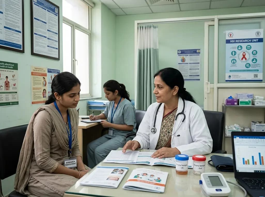
A secondary analysis of an HIV cure-related trial in South Africa highlights the acceptability and safety of serial excisional lymph node and gut biopsies among young women. Clear communication, empathetic care, and altruistic motives helped mitigate initial apprehension and sustain longitudinal trial engagement.
Today
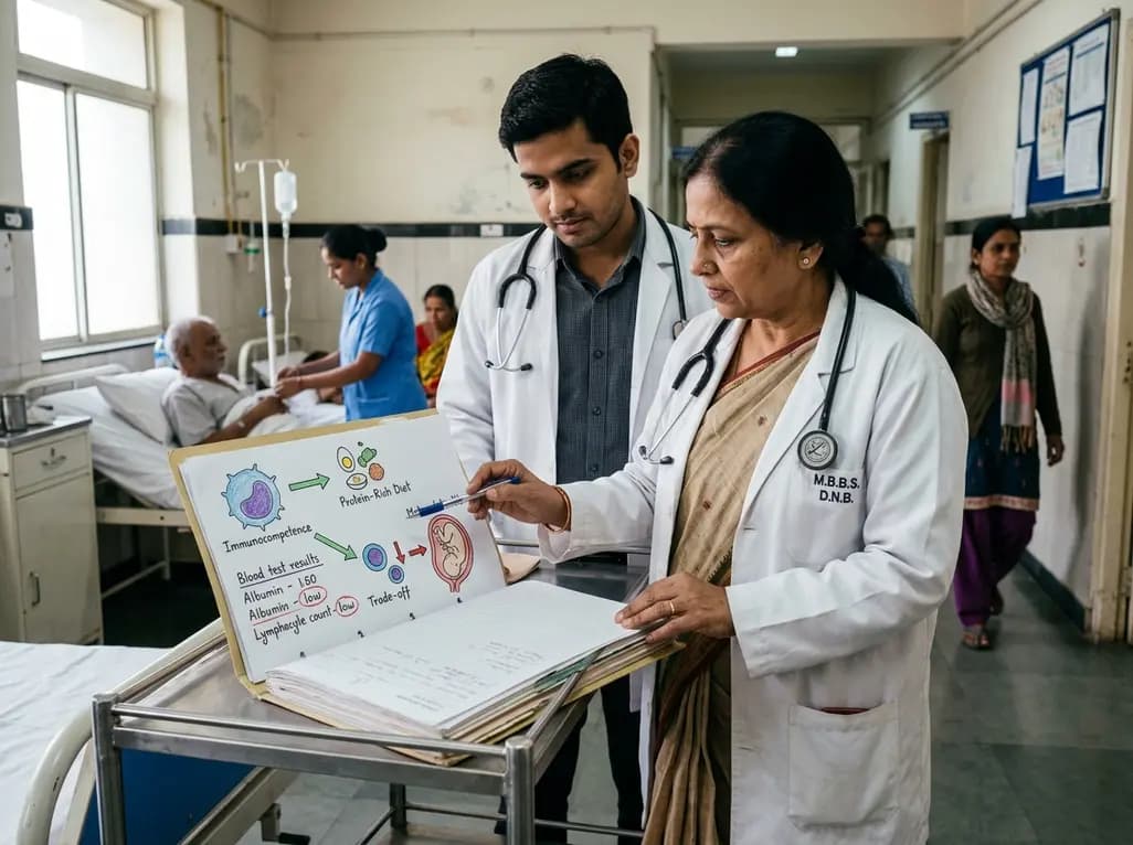
Explore the crucial link between dietary protein and immunocompetence. This review examines how low-protein nutrition shifts metabolic resources away from pathogen defense toward early reproduction, compromising host survival and immunity against severe bacterial infections.
Today
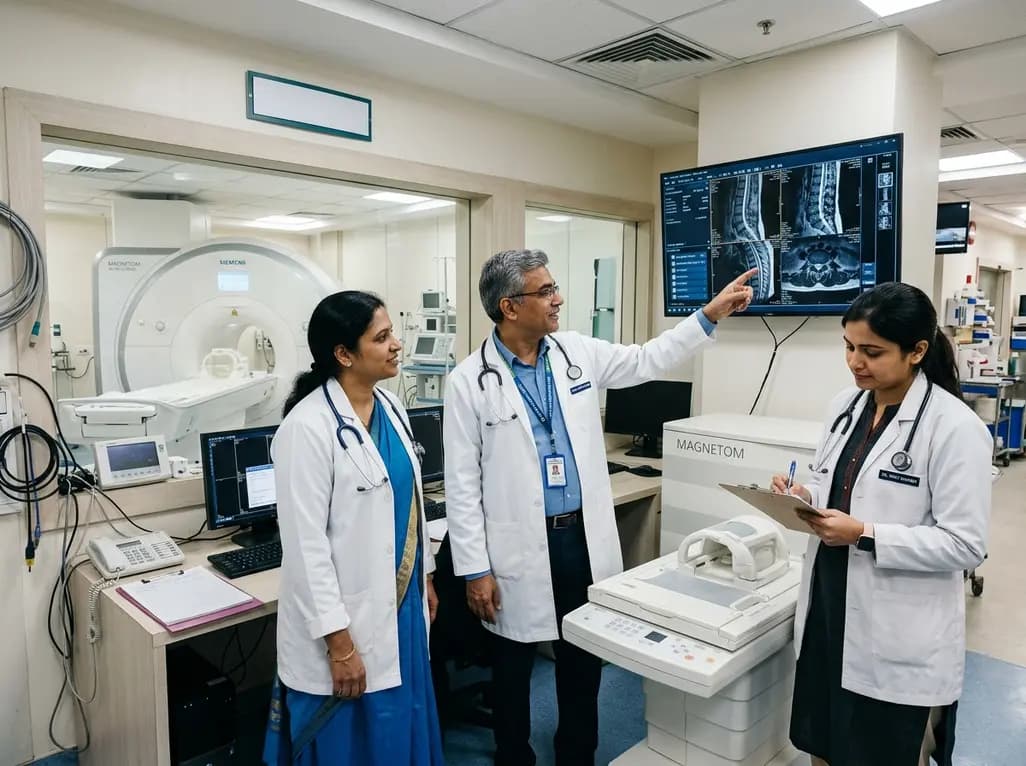
Under India's PLI initiative, domestic production of 57 critical medical devices has commenced, including MRI and CT scanners, cath labs, and heart valves. Global partnerships and local investments are expanding high-end technology, curbing import reliance, and strengthening clinical accessibility nationwide.
Today

A longitudinal study reveals that patients undergoing staged bilateral hip arthroscopy experience progressive contralateral labral deterioration during surgical intervals. However, structural degradation does not compromise 2-year postoperative clinical outcomes or patient-reported satisfaction scores.
Today
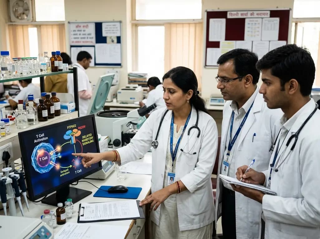
New immunological research reveals how PTPN22 forms a delayed inhibitory signaling complex to restrain T cell activation, explaining how the autoimmune-linked R620W variant disrupts feedback control and promotes persistent inflammation in conditions like rheumatoid arthritis and type 1 diabetes.
Today
