.webp&w=384&q=75)

Loading, please wait...
Loading, please wait...

Successful customized titanium mesh application relies heavily on precise surgical placement during guided bone regeneration (GBR). The anterior maxilla presents significant aesthetic and anatomical challenges for clinicians. Consequently, dental professionals are increasingly exploring digital workflows to improve predictable outcomes. This retrospective case series evaluated the effectiveness of using a vacuum-formed placing template to guide three-dimensionally printed mesh in ten patients requiring maxillary reconstruction.
Notably, researchers found that the customized titanium mesh demonstrated high graft volume accuracy, reaching approximately 91.1%. The team reconstructed cone-beam computed tomography (CBCT) data across three stages to measure displacement and contour discrepancies. Specifically, the mesh exhibited a minor buccal displacement of 0.57 mm immediately after surgery. Furthermore, the contour discrepancy decreased significantly by the five-to-eight-month follow-up period. These findings suggest that the vacuum-formed template provides a stable and feasible method for positioning the mesh accurately within the defect site.
However, mesh exposure remains a common clinical hurdle in bone augmentation. The study reported non-infected mesh exposure in 26% of implant sites. Additionally, vertical bone resorption varied significantly between groups. Exposed sites experienced a resorption of 1.69 mm, whereas non-exposed sites showed only 0.14 mm of loss. Therefore, maintaining soft tissue integrity is crucial for preserving the newly formed bone. Overall, this technique validates the utility of digital templates in achieving superior spatial maintenance for complex anterior defects.
The vacuum-formed template acts as a physical guide that aligns the 3D-printed mesh with the preoperative digital design. This tool reduces manual errors and ensures the mesh remains in the intended buccal and apical position during fixation.
While customized mesh is highly effective, exposure can lead to increased vertical bone resorption. Evidence suggests that non-exposed sites retain significantly more bone height compared to those where the mesh becomes visible during the healing phase.
Customized 3D-printed meshes offer a better anatomical fit and require less manual contouring during surgery. This precision helps in maintaining the space required for regeneration, particularly in complex defects where traditional mesh might collapse or shift.
Disclaimer: This content is for informational and educational purposes only. It is not intended as a substitute for professional medical advice, diagnosis, or treatment. Always seek the advice of your physician or other qualified health provider with any questions you may have regarding a medical condition. Refer to the latest local and national guidelines for clinical practice.
References
Zhang S et al. Placement Error and Bone Regeneration of Customized Titanium Mesh With Vacuum-Formed Placing Template for Anterior Maxillary Bone Defects: A Retrospective Case Series Study. Clin Oral Implants Res. 2026 Mar 13. doi: 10.1111/clr.70115. PMID: 41826268.
Tallarico M et al. Customized 3D-Printed Titanium Mesh Developed to Regenerate a Complex Bone Defect in the Aesthetic Zone: A Case Report. Materials (Basel). 2020;13(17):3874.
Li L et al. Research on the dimensional accuracy of customized bone augmentation combined with 3D-printing individualized titanium mesh: A retrospective case series study. Clin Implant Dent Relat Res. 2021;23(1):5-18.
"
Read summarized clinical updates, watch expert medical content, and earn CME certifications right from your smartphone.
Open your smartphone camera to scan and install.





A retrospective study validates the use of vacuum-formed templates for precise positioning of 3D-printed titanium mesh in anterior maxillary bone defects....
4 months ago
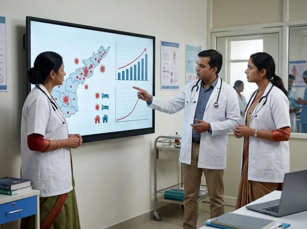
Andhra Pradesh reported 10 new Covid-19 cases, taking the state tally to 49 while deaths remain at four. With 24 patients hospitalized and 16 under home isolation, the Health Department has intensified monitoring. Medical professionals should review regional distribution, diagnostic protocols, and management plans.
Today

An 11-year Swedish registry study of 618 uterine sarcoma patients found that minimally invasive surgery yielded survival comparable to open surgery in early stages. However, adjuvant chemotherapy conferred no survival benefit in localized or advanced disease, highlighting stage and histology as key outcomes.
3 days back

A cross-sectional study evaluates post-intensive care syndrome in cardiac patients 2-4 weeks post-ICU discharge, highlighting cognitive, psychological, and functional impairments and the need for structured multidisciplinary rehabilitation.
3 days back

Anterior cruciate ligament reconstruction failure lacks uniform definition. A narrative review proposes an integrative framework incorporating objective and subjective instability, persistent pain, restricted motion, graft rupture, and secondary meniscal injury to standardize clinical reporting.
3 days back
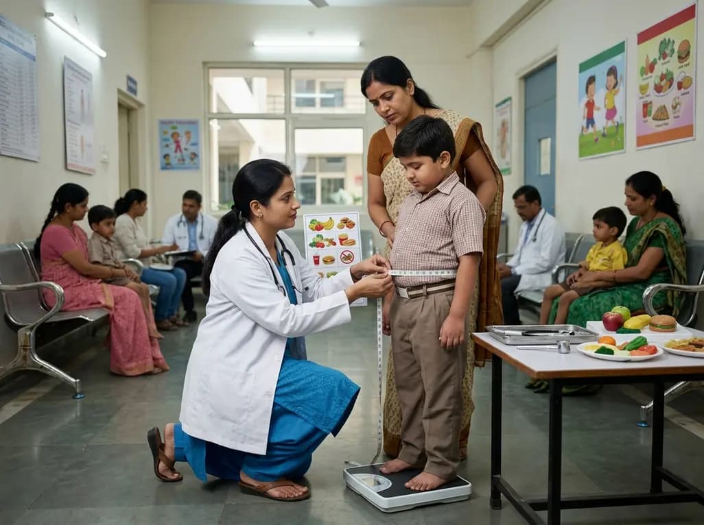
With World Obesity Atlas data warning that over 41 million Indian children are overweight or obese, ICMR and NIN have unveiled a 10-point policy roadmap. The initiative calls for mandatory front-of-pack labeling, HFSS taxes, strict marketing bans, and healthier school environments to curb non-communicable diseases.
Today
