.webp&w=384&q=75)

Loading, please wait...
Loading, please wait...

Superficial temporal artery to middle cerebral artery (STA-MCA) bypass remains the primary revascularization intervention for adult Moyamoya disease. However, neurosurgeons frequently encounter postoperative neurological deterioration triggered by cerebral hyperperfusion syndrome. This acute phenomenon occurs when restored arterial blood flow overwhelms chronically dilated, autoregulation-impaired intracranial microvessels. Consequently, the fragile capillary network develops focal hyperemia, severe vasogenic edema, and heightened intracerebral hemorrhage risk. Clinicians often observe focal neurological deficits, severe unilateral headaches, or acute seizures within the initial postoperative week. Furthermore, distinguishing hyperperfusion from acute graft failure or cerebral ischemia presents a profound diagnostic challenge. Misinterpreting these symptoms can prompt inappropriate hypertensive therapy that accelerates catastrophic hemorrhagic transformation. Therefore, neurosurgical teams urgently require reliable noninvasive predictive biomarkers to stratify patient risk before entering the operating theater. In addition, clinicians turned to advanced computed tomography perfusion (CTP) imaging to evaluate these precarious hemodynamic shifts. Despite enthusiastic clinical adoption, the actual prognostic power of perioperative perfusion parameters has remained uncertain across clinical cohorts. Specifically, clinicians must determine whether quantitative perfusion measurements can reliably detect tissue beds susceptible to hyperemic damage. Thus, establishing robust hemodynamic surveillance protocols represents a crucial objective in modern cerebrovascular surgery.
Moyamoya disease induces progressive, non-atherosclerotic stenosis of the terminal internal carotid arteries and their proximal branches. As a result, compensatory networks of fragile collateral vessels form to rescue ischemic brain parenchyma. Direct STA-MCA bypass effectively redirects extracranial blood directly into the ischemic middle cerebral artery territory. However, chronic hypoperfusion severely diminishes cerebral vascular reserve throughout vulnerable cerebral territories. Vascular smooth muscle cells in chronically ischemic zones maximize resting vasodilation, thereby completely exhausting their autoregulatory reserve capacity. Consequently, when the bypass delivers robust arterial flow, the paralyzed local vascular bed cannot constrict appropriately. This sudden surge produces focal hyperemia that breaches the fragile blood-brain barrier. In addition, the revascularized brain tissue can suffer transit time shifts, local tissue shearing, and microvascular extravasation. Clinicians must maintain a very delicate equilibrium between preventing bypass thrombosis and avoiding excessive microvascular pressure. Lowering blood pressure excessively risks ischemic stroke in collateral-dependent non-revascularized zones. Conversely, allowing elevated postoperative blood pressure rapidly fuels vasogenic edema and triggers life-threatening intracerebral hematomas. Therefore, understanding microvascular hemodynamics is vital for optimizing perioperative management. Moreover, neurointensivists continuously seek objective neuroimaging thresholds to identify patients who cannot tolerate sudden flow increases.
To clarify the diagnostic value of perfusion imaging, investigators conducted a rigorous systematic review and meta-regression following PRISMA guidelines. The researchers searched major biomedical databases, including MEDLINE and Embase, through July 2026. Specifically, the team examined adult patients with Moyamoya disease undergoing STA-MCA bypass revascularization procedures. Accordingly, the systematic analysis focused on relative computed tomography perfusion values, evaluating preoperative, postoperative, and differential parameter shifts. These analyzed metrics included relative cerebral blood flow, relative cerebral blood volume, relative mean transit time, and relative time-to-peak. From 177 identified citations, investigators reviewed 40 full-text articles and included five rigorous studies representing 379 surgical patients. Within this primary pooled cohort, exactly 23 hyperperfusion events occurred, establishing a pooled complication incidence of 4.9 percent. Furthermore, the investigators conducted an expanded sensitivity analysis incorporating 11 studies and 962 patients. This broader sensitivity cohort added dynamic susceptibility contrast magnetic resonance perfusion and transcranial color Doppler datasets. Researchers subsequently applied univariate random-effects meta-regression alongside multivariate modeling to test parameter associations. In addition, the authors scrutinized methodological variations among clinical centers to eliminate confounding procedural differences. Ultimately, this comprehensive analytical framework provided the most exhaustive evaluation of CT perfusion metrics in Moyamoya bypass surgery to date.
Surprisingly, the meta-regression revealed that no individual CT perfusion parameter demonstrated a statistically significant association with post-bypass hyperperfusion. Neither preoperative relative cerebral blood flow nor elevated baseline blood volume reliably separated vulnerable patients from uneventful surgical cases. Similarly, postoperative transit time accelerations and differential hemodynamic calculations failed to achieve predictive significance across the cohorts. Even within the expanded multivariate sensitivity analysis, perfusion values showed no reproducible diagnostic correlation with clinical hyperperfusion events. Several distinct biological and methodological factors explain this lack of predictive performance. First, substantial heterogeneity exists regarding how clinical centers define and classify hyperperfusion syndrome. Some institutions require objective radiologic hyperemia alone, whereas other protocols demand overt focal neurological deficits before confirming the diagnosis. Second, standard regional regions-of-interest on CT perfusion slices often miss microvascular flow fluctuations occurring near the anastomosis. Cortical revascularization creates extreme local flow gradients that whole-brain or territorial region-of-interest averaging simply dilutes. Third, systemic post-surgical variables strongly modify hemodynamic outcomes regardless of baseline scans. Therefore, resting static imaging metrics cannot fully capture the dynamic physiological stress of surgical revascularization. Consequently, surgeons cannot rely on conventional CT perfusion alone to guide clinical decisions.
Because imaging thresholds remain unproven, neurosurgeons and neurocritical care teams must maintain meticulous clinical vigilance. Postoperative care requires standardized, proactive management rather than passive dependence on single neuroimaging scans. Clinicians must institute continuous arterial blood pressure monitoring immediately after completing the bypass anastomosis. Many neurovascular protocols recommend maintaining systolic blood pressure below 130 to 140 millimeters of mercury in adult Moyamoya patients. However, teams must avoid profound hypotension that could compromise flow through fragile collateral pathways. In addition, routine neurological evaluations every hour facilitate early recognition of transient motor deficits, aphasia, or focal seizures. If a patient develops new neurological signs, clinicians should promptly obtain cross-sectional imaging to exclude acute intracerebral hemorrhage or graft occlusion. When hyperperfusion is suspected, aggressive pharmacologic blood pressure lowering and free radical scavenging agents can prevent permanent parenchymal injury. Furthermore, emerging intraoperative tools, such as microvascular Doppler flow probes and cortical hyperspectral imaging, may offer superior real-time insight into flow shifts. Ultimately, future research requires large, prospective patient-level registries utilizing standardized definitions of hyperperfusion. Until researchers establish validated predictive models, multidisciplinary vigilance remains the cornerstone of safe Moyamoya bypass surgery.
Cerebral hyperperfusion syndrome occurs when direct surgical revascularization restores robust blood flow into chronically hypoperfused vascular territories. Because longstanding ischemia impairs local microvascular autoregulation, the paralyzed cerebral arterioles cannot constrict against sudden flow increases. Consequently, excessive hydrostatic pressure breaches the blood-brain barrier, triggering vasogenic edema, seizures, and intracerebral hemorrhage.
Recent meta-regression evidence demonstrates that CT perfusion parameters cannot reliably predict hyperperfusion risk. Neither baseline blood flow, transit time delays, nor differential postoperative perfusion changes show significant associations with clinical events. Therefore, clinicians must not depend exclusively on perfusion imaging thresholds to identify high-risk surgical patients in routine practice.
Clinicians should implement continuous intra-arterial blood pressure monitoring immediately after surgery. Protocols typically maintain systolic blood pressure within individualized normotensive ranges, often below 130 to 140 millimeters of mercury, to curb microvascular pressure. However, care teams must avoid excessive hypotension, which threatens collateral circulation in non-revascularized ischemic brain regions.
Disclaimer: This content is for informational and educational purposes only... Refer to the latest local and national guidelines for clinical practice.
References

Read summarized clinical updates, watch expert medical content, and earn CME certifications right from your smartphone.
Open your smartphone camera to scan and install.





A meta-regression reveals that CT perfusion parameters do not reliably predict cerebral hyperperfusion syndrome following STA-MCA bypass in Moyamoya disease. Clinicians must rely on intensive perioperative blood pressure control and clinical monitoring rather than static imaging thresholds.
Today
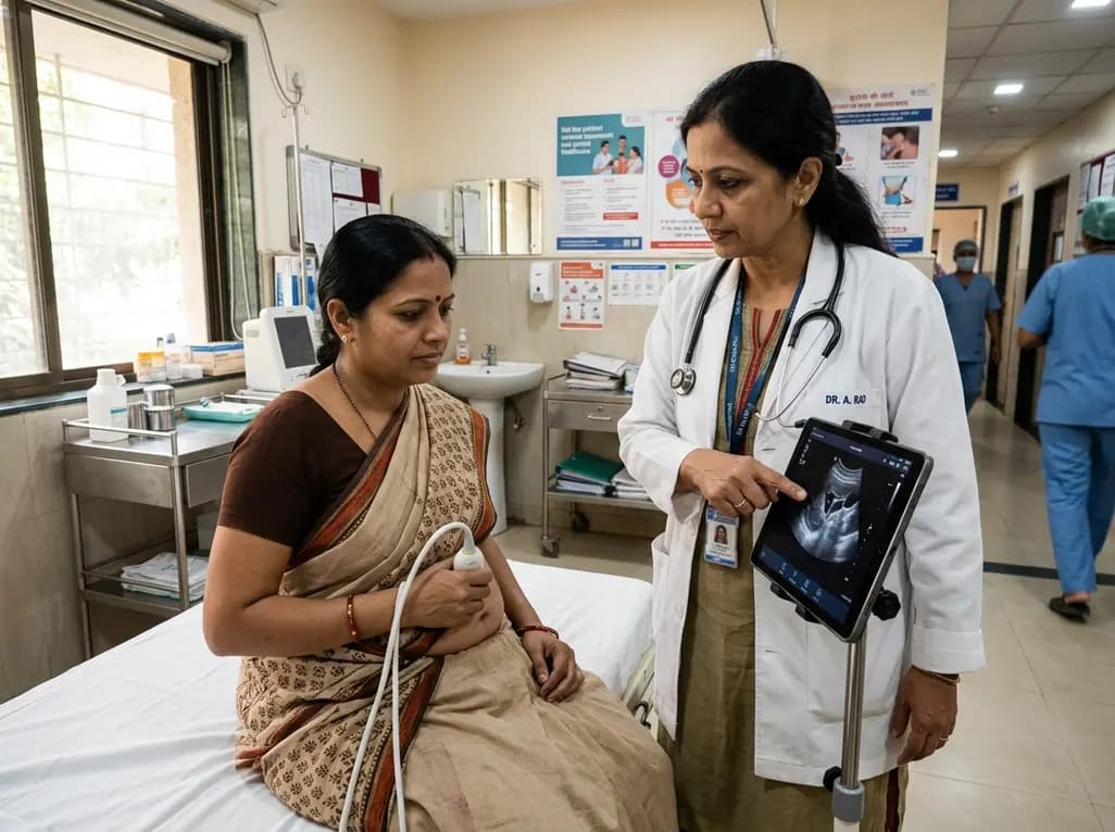
Self-performed ultrasound empowers patients to capture diagnostic-quality scans for remote clinical review. This review evaluates its accuracy, technological challenges, and integration hurdles.
Today
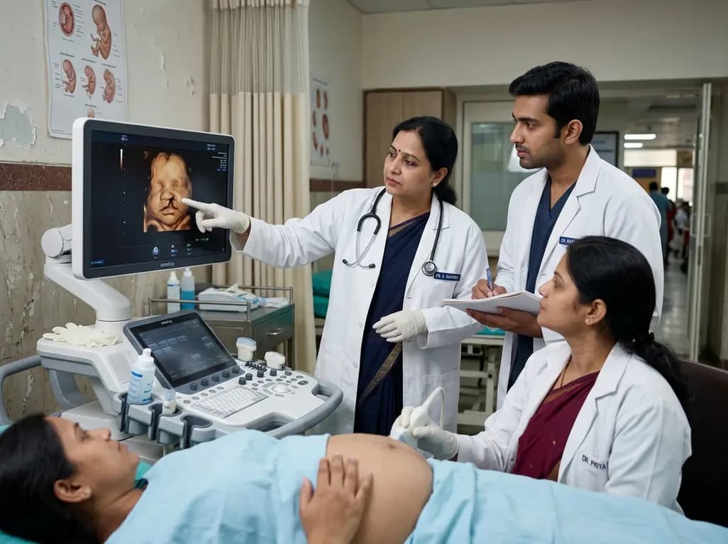
A 5-year retrospective study evaluates prenatal ultrasound detection of fetal cleft lip and palate. While overall detection reached 87%, accuracy varied from 99.3% in combined clefts to 50% in isolated cases. Combined defects showed elevated risks of concurrent structural anomalies and chromosomal abnormalities.
Today

An updated systematic review and meta-analysis of 7,875 patients reveals that routine microaxial left ventricular support during nonemergent high-risk PCI does not reduce mortality or ischemic complications, while significantly increasing major bleeding events.
Yesterday
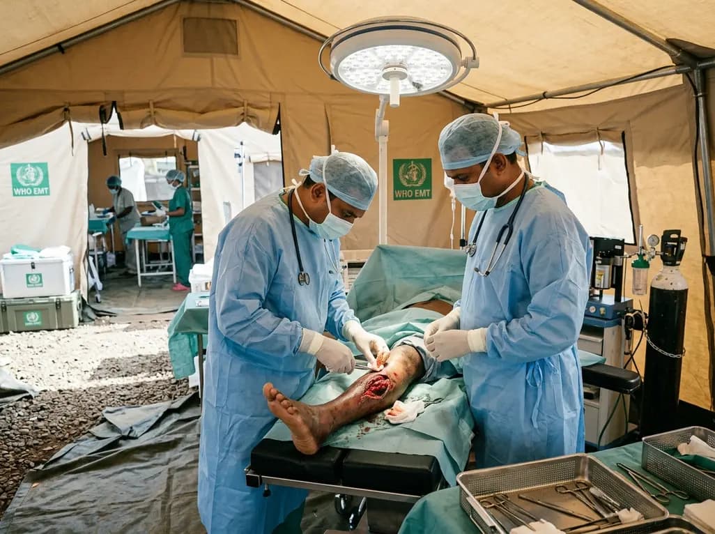
Following Cyclone Chido, a WHO EMT-2 field hospital performed 268 surgeries in Mayotte. While acute trauma is expected initially, 98% of cases were septic wounds of the extremities requiring secondary intention healing, revealing crucial operational insights for disaster surgical preparedness.
Today

The SED-SEEN-SEEP consensus outlines an Integrated Care Pathway for preclinical type 1 diabetes, structuring target screening, autoantibody confirmation, metabolic staging, and proactive follow-up to prevent diabetic ketoacidosis and facilitate early disease-modifying intervention.
Yesterday
