.webp&w=384&q=75)

Loading, please wait...
Loading, please wait...
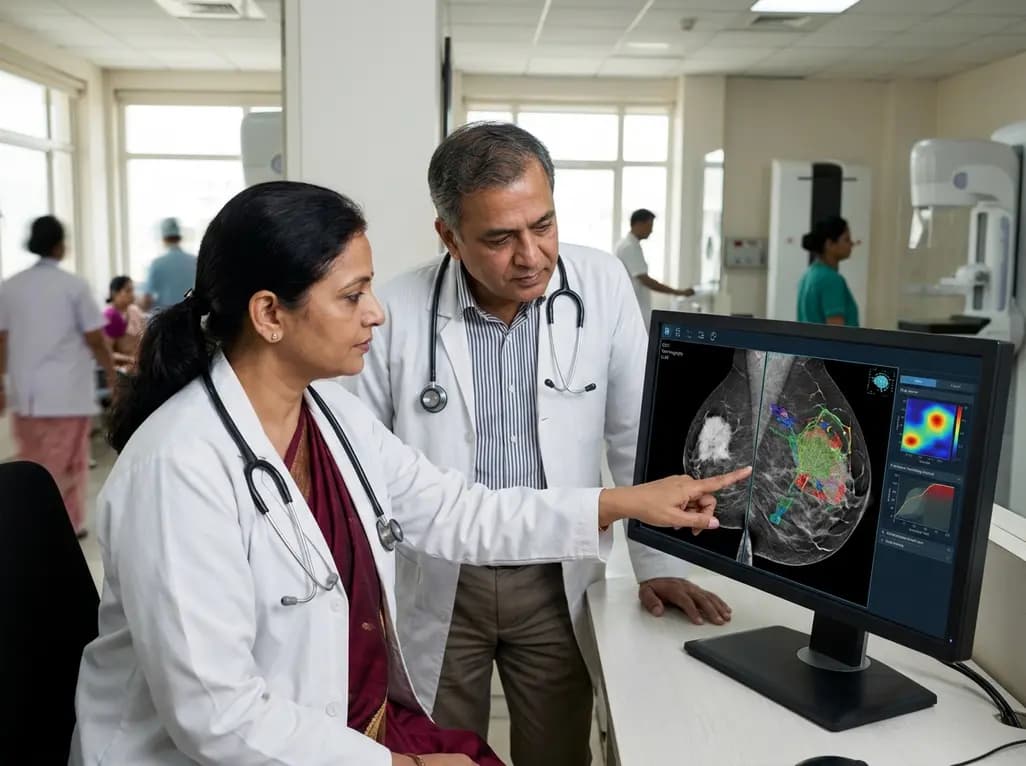
Accurate preoperative assessment is vital for managing patients with invasive breast cancer effectively. Recently, researchers have focused on contrast-enhanced mammography (CEM) as a powerful tool for predicting lymphovascular invasion (LVI) status. LVI is a critical prognostic marker that significantly influences surgical decisions and the need for adjuvant therapy. While traditional imaging often finds it difficult to identify LVI before surgery, CEM offers functional insights into tumor vascularity. This new research suggests that CEM can bridge the diagnostic gap, allowing for more personalized treatment planning.
Lymphovascular invasion involves the presence of cancer cells within the blood or lymphatic vessels. Its presence typically indicates a higher risk of nodal metastasis and systemic recurrence. Therefore, knowing the LVI status preoperatively helps surgeons determine the necessity of more aggressive axillary interventions. Furthermore, it aids oncologists in assessing the likely benefit of systemic treatments. In the Indian clinical context, where breast cancer is often diagnosed at later stages, such predictive tools are invaluable for optimizing resource use and improving patient outcomes.
The study analyzed 243 female patients who underwent preoperative contrast-enhanced mammography. Researchers examined several CEM features, including lesion enhancement patterns and background parenchymal enhancement (BPE). The results showed that specific features are significantly associated with LVI-positive status. For instance, lesions exhibiting complete enhancement or enhancement extending beyond the primary mass were strong indicators of invasion. Additionally, clinical factors like a high Ki67 index and axillary adenopathy were identified as independent predictors of LVI.
Beyond traditional statistical analysis, the study utilized six different machine learning methods to build predictive models. The LogitBoost model emerged as the top performer, achieving an Area Under the Curve (AUC) of 0.902 in the test dataset. This high level of accuracy demonstrates the potential for integrating artificial intelligence with CEM imaging. Consequently, clinicians can utilize these models to gain a clearer picture of a patient's risk profile before they even enter the operating room. Because CEM is often more accessible and cost-effective than MRI, it provides a practical solution for many healthcare settings in India.
The findings underscore the importance of a multimodal approach to breast cancer staging. Radiologists should pay close attention to enhancement characteristics on CEM, as these serve as surrogates for tumor aggressiveness. Meanwhile, surgeons can use this data to counsel patients more accurately regarding the extent of their upcoming procedures. By identifying high-risk patients early, the medical team can ensure that the most comprehensive care is provided from the outset.
CEM combines standard mammographic imaging with contrast medium to highlight areas of increased vascularity. This functional information allows for better detection of malignancies and more accurate assessment of tumor characteristics like LVI.
LVI is an independent risk factor for recurrence. Its presence often leads to a more aggressive treatment approach, which may include extensive axillary surgery or specialized chemotherapy regimens.
Yes, machine learning models like LogitBoost can analyze complex patterns in imaging data that may be subtle to the human eye. In this study, the model predicted LVI status with high accuracy, reaching an AUC of over 0.90.
Disclaimer: This content is for informational and educational purposes only. It is not a substitute for professional medical advice, diagnosis, or treatment. Always seek the advice of your physician or other qualified health provider with any questions you may have regarding a medical condition. Refer to the latest local and national guidelines for clinical practice.
References
Gong L et al. Use of contrast-enhanced mammography for preoperative prediction of lymphovascular invasion status in invasive breast cancer. Cancer Imaging. 2026 Jun 06. doi: 10.1186/s40644-026-01065-1. PMID: 42251446.
"
Read summarized clinical updates, watch expert medical content, and earn CME certifications right from your smartphone.
Open your smartphone camera to scan and install.





A study demonstrates that contrast-enhanced mammography (CEM) features and machine learning models can preoperatively predict lymphovascular invasion (LVI)....
4 months ago
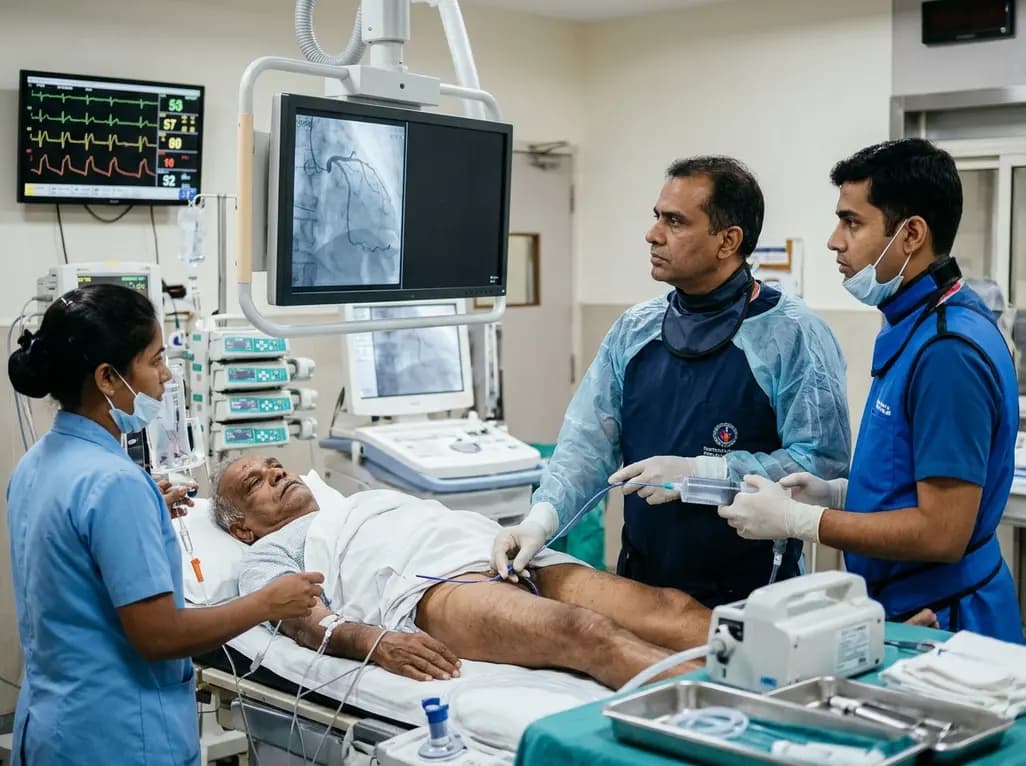
An elderly man with partially repaired Tetralogy of Fallot presented with STEMI caused by coronary embolism rather than atherosclerosis. Successful treatment with sustained mechanical aspiration thrombectomy underscores the vital need for high clinical vigilance in adult congenital heart disease.
Today

Demanding exhaustive hormonal profiling can inadvertently discourage clinical studies involving females. Methodological standards must align with the specific scientific question to expand, not restrict, equitable evidence for women.
Today
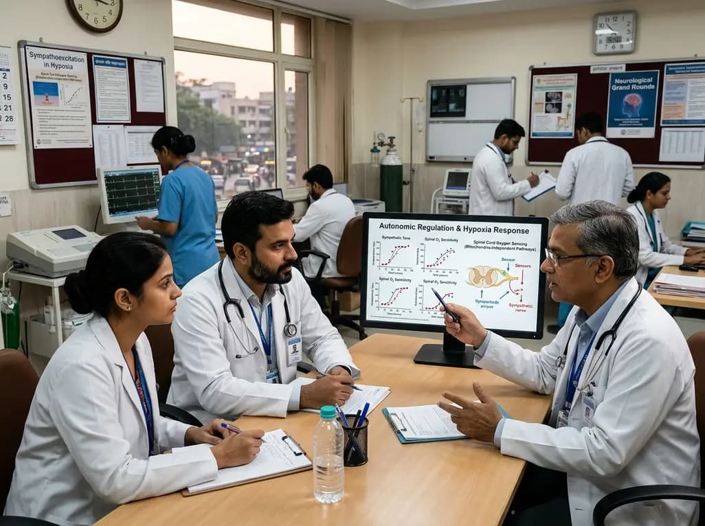
A recent study demonstrates that spinal oxygen sensors do not require mitochondrial electron transport to trigger sympathetic excitation during acute hypoxia, revealing new insights into how mitochondrial oxidative stress modulates autonomic tone in cardiovascular and neurological diseases.
Today

A novel calcium polyphosphate coacervate enables injectable bone implants with in situ hierarchical porosity via phase separation and gas foaming, significantly enhancing cell proliferation, energy metabolism, and osteogenic differentiation for minimally invasive bone defect repair.
Today

Following regulatory scrutiny from FSSAI regarding promotional representations and lean muscle preservation claims, Dr. Reddy's Laboratories and Nestlé Health Science are currently reviewing the branding and marketing strategy for their joint nutritional supplement Celevida GLP+.
Today
