.webp&w=384&q=75)

Loading, please wait...
Loading, please wait...

Central nervous system neoplasms represent the leading cause of disease-related mortality among children globally. Pediatric oncologists have long hypothesized that embryological aberrations contribute to oncogenesis. However, evaluating specific co-occurrences has historically remained challenging due to the relative rarity of individual neoplasms. A landmark population-based registry linkage study published in Neuro-Oncology provides unprecedented clarity on this question. Researchers examined medical registries covering nearly twenty-three million live births across nine states to evaluate the relationship between congenital malformations and childhood brain tumors. Their comprehensive findings establish clear histological associations and emphasize a critical vulnerability window during infancy.
To overcome statistical power limitations inherent in pediatric oncology cohorts, the investigators assembled data from 22,599,099 live births between 1990 and 2018. Consequently, this immense cohort enabled rigorous tracking of birth defect registries linked directly with regional cancer registries. The researchers identified 6,247 children diagnosed with primary central nervous system neoplasms before age twenty. Crucially, investigators classified anomalies into major structural defects without known chromosomal syndromes, chromosomal conditions, neurofibromatosis, and tuberous sclerosis complex. Additionally, the team applied multivariable Cox proportional hazards models adjusting for maternal age, maternal education, race, and birth state. Because birth registries documented defects prospectively, the investigation effectively eliminated recall bias. Furthermore, the protocol evaluated 66 distinct anomaly-tumor combinations that had at least five co-occurring events. Notably, 42 of these combinations achieved statistical significance, including 16 completely novel correlations that earlier small cohorts failed to capture. These robust methods provide pediatric healthcare providers with unprecedented epidemiological precision regarding developmental oncology risks.
The study revealed that the magnitude of risk diverges sharply depending on cancer histology. Overall, children born with any major structural anomaly experienced an elevated likelihood of developing childhood central nervous system malignancies, with adjusted hazard ratios spanning from 1.48 to 3.69. In particular, structural malformations correlated strongly with astrocytomas, atypical teratoid/rhabdoid tumors, medulloblastomas, and primitive neuroectodermal tumors. However, ependymomas exhibited no statistically significant association with nonchromosomal structural defects. This intriguing divergence strongly suggests that distinct oncogenic pathways govern ependymoma pathogenesis compared to other embryonal neoplasms. Furthermore, the investigators observed that risks differed substantially across astrocytoma histologies. For instance, children with specific structural anomalies displayed distinct patterns between low-grade pilocytic astrocytomas and high-grade malignant lesions. Therefore, clinicians must recognize that congenital anomalies do not exert a uniform oncogenic influence across all brain regions. Instead, early developmental perturbations appear to target specific cell lineages, altering neuroepithelial differentiation and driving lineage-dependent oncogenesis.
Age stratification provided some of the most striking insights in this large population-based linkage analysis. Specifically, the association between major structural defects and neuro-oncological disease peaked during early infancy. Children diagnosed before their first birthday demonstrated the highest relative risk, with hazard ratios exceeding 5.0 for several tumor categories. In contrast, relative risks attenuated steadily among older children, adolescents, and young adults. This pronounced temporal pattern suggests that prenatal disruptions accelerate oncogenesis early in postnatal life. Moreover, multiple congenital malformations further compounded this vulnerability. Children harboring two or more major structural anomalies exhibited markedly higher tumor hazard ratios than those with isolated defects. Consequently, clinicians evaluating infants with complex syndromic presentations must appreciate this heightened early-life risk profile. While the absolute risk remains modest, early neurological signs in this vulnerable subset warrant timely evaluation. Recognizing that the highest vulnerability concentrates in the first year of life enables pediatricians to calibrate their diagnostic vigilance appropriately.
The co-occurrence of structural birth defects and pediatric cancer points toward common embryological mechanisms. During embryogenesis, delicate signaling cascades regulate cellular proliferation, spatial migration, and terminal tissue differentiation. Therefore, disruptions in critical developmental pathways—such as Sonic Hedgehog, WNT, and MAP kinase cascades—can perturb organogenesis while simultaneously priming stem cell niches for malignant transformation. For instance, genes controlling cerebellar development and neural tube closure directly interact with pathways implicated in medulloblastoma formation. Similarly, early disruptions in chromatin remodeling complexes like SMARCB1 produce both structural renal or neural malformations and aggressive rhabdoid tumors. Furthermore, shared environmental teratogens or parental epigenetic alterations may concurrently induce abnormal structural morphogenesis and genomic instability. Consequently, clinicians should not view abnormal organogenesis as an isolated mechanical defect. Rather, physicians should consider structural malformations as phenotypic markers of broader germline or early postzygotic dysregulation. These mechanistic intersections offer vital biological clues for laboratory investigators studying the fetal origins of pediatric central nervous system malignancies.
Although these epidemiological associations are striking, pediatric clinicians must translate them into measured, evidence-based bedside practice. First, healthcare providers should reassure parents that primary brain tumors remain rare in absolute numbers, even among children born with congenital malformations. As a result, routine magnetic resonance neuroimaging is currently unjustified for asymptomatic children with isolated structural defects. Such widespread screening would generate excessive false positives, unnecessary contrast exposures, and heightened parental anxiety. Instead, physicians should emphasize targeted clinical vigilance and longitudinal developmental assessments. Specifically, neonatologists and pediatricians must maintain a low threshold for diagnostic neuroimaging when infants with congenital anomalies manifest neurological warning signs. Red flags include rapid head circumference growth, persistent unexplained vomiting, cranial nerve palsies, focal motor deficits, or significant developmental regression. Additionally, comprehensive clinical genetics evaluations remain essential for identifying underlying syndromes like neurofibromatosis or tuberous sclerosis. Ultimately, integrating structural anomaly awareness into routine pediatric care ensures earlier detection while preventing unwarranted radiological intervention.
Epidemiological data indicate that major structural birth defects increase tumor risk across most histological types, yielding adjusted hazard ratios between 1.48 and 3.69. Furthermore, infants presenting with congenital defects exhibit even higher relative risks. Consequently, these robust findings suggest a shared biological susceptibility originating early during fetal morphogenesis.
The strongest co-occurrences emerge among embryonal tumors, atypical teratoid/rhabdoid tumors, and specific astrocytoma histologies. Conversely, researchers observed no statistically significant association between major structural congenital anomalies and ependymoma. Therefore, embryological disruptions appear to influence central nervous system oncogenesis selectively based on the cell lineage involved.
Currently, routine radiological screening remains unfeasible because the absolute baseline risk of pediatric central nervous system malignancies stays extremely low. Nevertheless, clinicians should maintain elevated clinical vigilance. Specifically, pediatricians must promptly investigate persistent neurologic deficits, macrocephaly, intractable vomiting, or unexplained developmental regressions in children born with significant structural anomalies.
Disclaimer: This content is for informational and educational purposes only... Refer to the latest local and national guidelines for clinical practice.
References

Read summarized clinical updates, watch expert medical content, and earn CME certifications right from your smartphone.
Open your smartphone camera to scan and install.





A landmark population-based study of over 22 million live births demonstrates a significant association between major structural congenital anomalies and childhood brain tumors, with the highest relative risks observed during infancy and varying across distinct tumor histologies.
Today

A North Texas pilot study reveals that combining virtual physician consultations with in-person house calls safely manages adult pneumonia. By utilizing CRB-65 severity scores, clinicians effectively triaged patients, providing home-based recovery while directing higher-risk individuals to emergency care.
Today

A quasi-experimental study demonstrates that culturally tailored educational interventions significantly boost HPV knowledge and vaccination intentions. These findings provide actionable strategies for healthcare providers in India to overcome vaccine hesitancy and advance cervical cancer prevention.
Today

A comprehensive meta-analysis evaluates the association between Body Mass Index and outcomes following abdominal aortic aneurysm repair. Discover how overweight and obesity correlate with lower mortality while underweight status confers heightened risk, challenging conventional preoperative risk stratification.
Today

The SOLARIO trial shows that a short course (≤7 days) of systemic antibiotic therapy is noninferior to long courses (≥4 weeks) in orthopedic infections treated with local antibiotic carriers, with significantly fewer adverse events.
Today
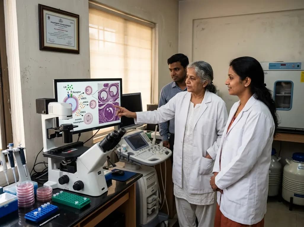
New research reveals that preovulatory follicles communicate via LH-induced paracrine signaling. Diffusible EGF ligands epiregulin and amphiregulin coordinate oocyte meiotic resumption across neighboring follicles, challenging traditional concepts of follicular autonomy.
Today
