

Loading, please wait...
Loading, please wait...

Adult neuro-oncology requires precise diagnostic classification to formulate effective surgical and medical strategies. For patients presenting with suspected or histologically confirmed grade II diffuse glioma, accurate baseline imaging dictates timely intervention. Conventional magnetic resonance imaging, however, frequently presents interpretive dilemmas when separating low-grade tumors from malignant lesions. To resolve these diagnostic ambiguities, the Congress of Neurological Surgeons published an updated systematic review and evidence-based guideline. This critical document establishes standardized pathways for advanced physiological neuroimaging and molecular tracer applications in routine neurosurgical practice.
Accurate baseline characterization of adult grade II diffuse glioma remains essential for effective clinical decision-making. Conventional magnetic resonance sequences, including T2-weighted and pre- and post-contrast T1-weighted acquisitions, serve as the historical benchmark for tumor evaluation. However, non-enhancing infiltrating margins frequently conceal aggressive tumor biology. Standard anatomical sequences often fail to identify focal anaplastic transformation within an apparent low-grade neoplasm. Consequently, clinicians may underestimate tumor aggressiveness, which can lead to delayed intervention or incomplete surgical planning.
Furthermore, post-treatment alterations introduce substantial diagnostic confusion during routine longitudinal surveillance. Radiation injury, inflammatory changes, and pseudoprogression closely simulate genuine disease recurrence on standard anatomical scans. Therefore, clinicians require advanced physiological parameters to distinguish indolent tumor margins from active cellular proliferation. Relying solely on conventional structural imaging compromises long-term diagnostic accuracy. In contrast, incorporating advanced physiological modalities enables neuro-oncology teams to delineate active disease early and avoid unnecessary exploratory procedures.
The updated guidelines establish clear recommendations regarding advanced physiologic magnetic resonance sequences. Specifically, the panel issued a Level II recommendation supporting diffusion-weighted imaging alongside perfusion-weighted imaging. These modalities encompass dynamic susceptibility contrast, dynamic contrast enhancement, and arterial spin labeling sequences. Clinicians should utilize these techniques whenever conventional structural sequences cannot definitively distinguish a low-grade tumor from high-grade glioma.
Perfusion sequences quantify relative cerebral blood volume, providing direct physiological insight into tumoral neoangiogenesis. In contrast, diffusion imaging assesses tissue cellularity by tracking extracellular water movement through apparent diffusion coefficient maps. Thus, elevated microvascular perfusion and restricted diffusion strongly suggest higher-grade biological behavior, even without overt contrast enhancement. Moreover, arterial spin labeling offers completely non-invasive vascular quantification without requiring gadolinium contrast administration. Consequently, neurosurgeons can reliably identify high-risk intratumoral zones, which facilitates targeted stereotactic biopsies and optimizes maximal safe surgical resection margins.
Magnetic resonance spectroscopy provides non-invasive metabolic profiling of intracranial neoplasms. Under the updated CNS guidelines, the panel assigned a Level III recommendation for proton magnetic resonance spectroscopy. Clinicians should consider spectroscopic analysis when standard anatomical sequences, diffusion techniques, and perfusion parameters fail to establish tumor grade definitively.
Spectroscopic evaluations quantify critical cellular metabolites, including choline, creatine, N-acetylaspartate, and myo-inositol. Actively proliferating glial neoplasms display elevated choline peaks reflecting accelerated membrane turnover. Concurrently, these lesions exhibit decreased N-acetylaspartate signals due to loss of healthy neuronal tissue. Therefore, an elevated choline-to-creatine or choline-to-N-acetylaspartate ratio indicates high cellular proliferation. Furthermore, specialized spectral editing sequences detect 2-hydroxyglutarate, which indicates isocitrate dehydrogenase gene mutations. Consequently, magnetic resonance spectroscopy supplies non-invasive biochemical validation that aids individualized diagnostic stratification when conventional scans yield inconclusive results.
Beyond advanced magnetic resonance techniques, nuclear medicine offers valuable molecular imaging modalities. The updated guideline provides Level III recommendations for amino acid positron emission tomography using fluoroethyl-L-tyrosine and fluorodopa tracers. Standard fluorodeoxyglucose PET displays intense physiological uptake within normal cerebral cortex, which obscures infiltrating glial margins. In contrast, radiolabeled amino acid tracers maintain minimal baseline accumulation in healthy brain tissue.
Consequently, amino acid tracers generate exceptional lesion-to-background contrast across infiltrative parenchymal boundaries. The guidelines suggest adding fluoroethyl-L-tyrosine or fluorodopa PET when magnetic resonance findings remain ambiguous regarding histological grade. In addition, novel amino acid PET metrics assist clinicians in estimating individual patient prognosis. Moreover, fluorodopa PET demonstrates exceptional sensitivity for distinguishing genuine tumor progression from radiation-induced necrosis. Incorporating these molecular radiotracers refines diagnostic certainty, especially when managing anatomically complex or diffusely infiltrative neoplasms.
The updated recommendations highlight the expanding role of advanced imaging in molecular characterization and surveillance. The expert panel established a Level III recommendation supporting diffusion and perfusion imaging to gather information regarding genomics and long-term prognosis. In modern neuro-oncology, molecular status drives therapeutic classification and prognostic stratification.
Advanced neuroimaging sequences correlate closely with key molecular markers, including IDH mutation status and 1p/19q codeletion. For instance, oligodendrogliomas frequently exhibit distinct perfusion profiles and vascular patterns on arterial spin labeling compared to astrocytomas. Furthermore, serial surveillance using advanced physiologic sequences detects malignant transformation before gross anatomical changes emerge. Therefore, regular physiologic monitoring allows oncologists to adjust adjuvant regimens proactively. By identifying rising perfusion metrics or declining apparent diffusion coefficient values, clinicians intervene well before irreversible clinical deterioration occurs.
Translating these evidence-based imaging guidelines into daily hospital practice demands seamless multidisciplinary coordination. Neurosurgeons, neuroradiologists, neuropathologists, and medical oncologists must formulate unified local imaging protocols. Hospital systems should standardize standardized low-grade glioma imaging templates across all high-field magnetic resonance scanners. Standardizing sequences ensures reproducible longitudinal comparisons across sequential follow-up scans.
Additionally, centers must expand access to advanced post-processing software for quantitative perfusion and diffusion analysis. Clinicians should review ambiguous neuroimaging studies within multidisciplinary tumor boards before altering surgical or systemic management. Furthermore, teams should consider amino acid PET whenever conventional imaging contradicts clinical presentation. Adopting this integrated framework minimizes diagnostic delays, optimizes targeted tissue sampling, and avoids unnecessary repeat surgeries. Ultimately, adhering to structured neuroimaging guidelines improves functional preservation and clinical outcomes for every adult patient.
Clinicians should order diffusion and perfusion sequences when standard T1- and T2-weighted MRI scans fail to establish histological tumor grade definitively. These advanced physiological sequences detect microvascular proliferation and hypercellularity, which helps differentiate lower-grade gliomas from aggressive high-grade malignancies before surgical planning.
Amino acid tracers, such as fluoroethyl-L-tyrosine and fluorodopa, provide superior contrast compared to standard FDG-PET because healthy brain tissue exhibits low baseline uptake. Consequently, these tracers clarify indeterminate tumor grades, define infiltrative margins, assist in prognostic stratification, and differentiate active tumor recurrence from treatment-related necrosis.
Yes, perfusion and diffusion parameters correlate significantly with underlying molecular genomics, including IDH mutation and 1p/19q codeletion profiles. While histological and molecular tissue analysis remains the definitive standard, advanced physiologic imaging provides valuable preliminary genomic and prognostic insights when comprehensive tissue testing is delayed or unavailable.
Disclaimer: This content is for informational and educational purposes only... Refer to the latest local and national guidelines for clinical practice.
References
Badve C et al. Congress of neurological surgeons systematic review and evidence-based guidelines for the role of imaging in newly diagnosed WHO grade II diffuse glioma in adults: update. J Neurooncol. 2025 Aug. doi: 10.1007/s11060-025-05043-8. PMID: 40338482.
Ormond DR et al. Congress of Neurological Surgeons Systematic Review and Evidence-Based Guidelines for the Treatment of Adults With WHO Grade II Diffuse Glioma: Update. Neurosurgery. 2026 May 01;98(5):984-991. doi: 10.1227/neu.0000000000003912. PMID: 41983690.
Olson JJ et al. Congress of Neurological Surgeons systematic review and evidence-based guidelines on the management of recurrent diffuse low-grade glioma: update. J Neurooncol. 2025 Aug;174(1):1-5. doi: 10.1007/s11060-025-05042-9. PMID: 40338481.

Read summarized clinical updates, watch expert medical content, and earn CME certifications right from your smartphone.
Open your smartphone camera to scan and install.





The Congress of Neurological Surgeons updated its evidence-based guidelines on neuroimaging for adult WHO grade II diffuse glioma. The recommendations clarify the roles of advanced diffusion MRI, perfusion sequences, MR spectroscopy, and amino acid PET tracers like FET and FDOPA in grading and surveillance.
Today

A cross-sectional molecular epidemiological study evaluates HIV-1 transmission clusters and pretreatment drug resistance among treatment-naïve men who have sex with men. Findings highlight prominent CRF07_BC transmission and significant assortative mixing, informing targeted public health and antiretroviral strategies.
Today

A new study evaluates a multilingual digital health platform combining rule-based cervical cancer risk assessment, retrieval-augmented generation AI, and geospatial navigation. The tool dramatically reduces clinical hallucinations while providing actionable health guidance for underserved populations.
Today

A novel MOF-based platform encapsulates hemoglobin with catalytic platinum and ceria nanozymes to deliver oxygen while scavenging toxic reactive oxygen species. This study highlights key design principles for managing hypoxia and reperfusion injury while resolving crucial stability challenges.
Today
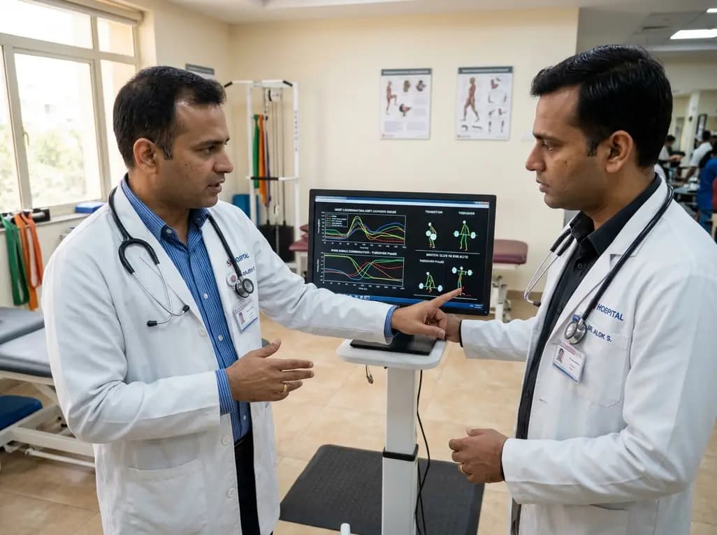
A vector coding study compares elite and sub-elite weightlifters during maximal snatch attempts, revealing that performance differences stem from phase-dependent knee-ankle coordination during transition and turnover phases rather than overall trajectory patterns.
Today

A pilot feasibility study demonstrates that a newly developed mobile app for bolus insulin dose adjustment significantly improves dosing confidence, treatment adherence, and hypoglycemia prevention for patients with type 1 diabetes while reducing provider workload in resource-limited clinical settings.
Today
