.webp&w=384&q=75)

Loading, please wait...
Loading, please wait...

Cervical malignancies do not develop overnight, but rather progress through slow cellular changes that can persist unnoticed for several years. Fortunately, researchers are now designing sophisticated algorithms to detect these subtle warning signs long before invasive tumors can form. Specifically, a groundbreaking study from Bengaluru demonstrates how advanced machine learning models can revolutionize early diagnostic protocols. At Christ University, researcher Lalasa Mukku has pioneered a suite of innovative artificial intelligence models. These systems identify patients at elevated risk by analyzing precancerous changes. These subtle lesions are clinically known as Cervical Intraepithelial Neoplasia. Consequently, the research group patented a predictive platform designed to forecast cancer risk five years prior to tumor manifestation. This proactive diagnostic timeline offers a critical window for preventive care. Therefore, integrating cervical cancer AI screening into routine practice can shift clinical focus from treatment to early prevention. By combining advanced computer vision with clinical files, clinicians can identify high-risk individuals earlier than traditional protocols allow. Ultimately, this paradigm shift promises to decrease mortality rates and redefine women's healthcare across diverse clinical environments. Medical professionals can utilize these predictions to schedule timely interventions and save lives.
Cervical intraepithelial neoplasia represents the premalignant phase of cervical carcinoma, which demands early clinical detection. Globally, cervical cancer remains one of the leading causes of death among women. Unfortunately, a vast majority of these preventable deaths occur in low- and middle-income nations. In these regions, access to specialized healthcare and comprehensive screening infrastructure is extremely limited. Consequently, many patients present with advanced disease stages, which significantly reduces their survival rates. However, identifying precancerous lesions during early screening can drastically improve clinical outcomes. Clinicians typically perform colposcopy examinations to inspect the cervix for abnormal cellular patterns. During these visual tests, gynecologists apply saline, acetic acid, and iodine solutions to highlight tissue variations. Each solution highlights the cellular landscape differently, creating distinct visual markers [1]. Therefore, precise interpretation of these sequential changes is critical to identify early dysplasia. By using machine learning to interpret these stages, medical providers can standardize diagnosis and eliminate subjective bias. Furthermore, this digital approach bridges the gap between rural health centers and expert diagnostic services. Ultimately, timely detection allows clinicians to intervene with minimally invasive therapies before invasive cancer develops.
To enhance diagnostic precision, Lalasa Mukku developed an advanced algorithm named the Colposcopic Multimodal Temporal Convolution Neural Network. This model combines sequential images from colposcopy with the patient’s structured clinical data. Specifically, the system processes visual inputs captured after applying saline, acetic acid, and iodine solutions. Each stage provides distinct physiological details, which are then fused with clinical histories. In a key 2024 study, this model demonstrated outstanding performance [1]. Consequently, the CMT-CNN model achieved an impressive classification accuracy of 92.3% in identifying cervical intraepithelial neoplasia. This multimodal approach outperforms classical single-image analysis by analyzing temporal changes across different clinical stages. Moreover, the integration of patient medical records provides critical context that raw images alone cannot supply. Clinicians can leverage this objective computer assistance to make highly informed decisions during patient evaluations. Thus, the model serves as an intelligent decision-support tool in busy clinical environments. Additionally, this technology can significantly reduce the workload of specialized pathologists in low-resource settings. By streamlining the screening process, the algorithm ensures that high-risk cases receive immediate specialist attention. Ultimately, this framework establishes a new standard for automated screening.
Despite the high potential of deep learning, analyzing colposcopic images presents unique technical challenges. For instance, moisture on the cervix surface often causes bright specular reflections during examinations. These reflections resemble white lesions, which are key clinical indicators of precancerous changes [1]. Consequently, standard computer vision models frequently misinterpret these reflections, leading to false-positive results [1]. To address this issue, Mukku developed a separate, highly specialized image-processing technique [1]. Specifically, this innovative method automatically detects and removes specular reflections prior to core analysis. Furthermore, the preprocessing pipeline isolates the cervix region with high structural accuracy [1]. As a result, the subsequent classification algorithm focuses exclusively on true cellular lesions without background noise. Importantly, this preprocessing step significantly increases the overall reliability and accuracy of automated cervical diagnostics. Therefore, clinicians can trust the machine's findings with much greater confidence. In addition, removing these visual artifacts reduces the need for repeating colposcopies, saving valuable time and clinical resources. This technical breakthrough directly translates to better patient experiences and optimized workflows. Ultimately, solving this image quality issue represents a vital milestone in deploying AI tools into real-world hospital environments.
In her latest research, Mukku introduced a cutting-edge quantum convolutional neural network to analyze complex medical images [1]. Specifically, she proposed this innovative architecture at an international IEEE conference in 2025 [1]. This advanced system leverages quantum computing principles to execute complex feature extractions much faster than classical deep networks. To test its diagnostic efficacy, the research team evaluated the model on public cervical screening datasets [1]. Remarkably, the quantum-inspired model achieved an extraordinary classification accuracy of approximately 98.6% [1]. This exceptional rate clearly demonstrates the immense potential of quantum computing in modern healthcare. Furthermore, quantum networks can process large, high-dimensional patient datasets with minimal computational overhead. This dramatic efficiency makes them highly suitable for implementation in resource-constrained healthcare facilities. Consequently, local providers can obtain near-instantaneous risk assessments during standard patient consultations. However, this technology currently remains in the research phase and requires extensive clinical validation [1]. Researchers must perform rigorous clinical trials across diverse populations before hospital deployment [1]. Nevertheless, this work paves the way for highly accurate, scalable cancer screening solutions globally. Ultimately, combining quantum mechanics with artificial intelligence could redefine the future of clinical oncology.
Q1: What is the CMT-CNN model and how does it detect precancerous changes?
The Colposcopic Multimodal Temporal Convolution Neural Network is an AI-based system designed to classify cervical intraepithelial neoplasia. Specifically, the system processes sequential images captured during colposcopy after applying saline, acetic acid, and iodine solutions. It fuses these temporal visual changes with the patient's structured clinical data. Consequently, the model achieved 92.3% accuracy, providing critical risk assessments years before tumor formation.
Q2: How does the new image-processing technique handle specular reflections on the cervix?
Moisture on the cervix surface often causes bright specular reflections during examinations, which look like precancerous lesions [1]. To address this, the researcher developed a separate image-processing algorithm that automatically removes these reflections [1]. Additionally, this technique isolates the cervical region with high precision before core machine learning analysis [1]. As a result, this pipeline improves diagnostic reliability, prevents false-positive interpretations, and minimizes repeated procedures for patients.
Q3: What are the main benefits of using a quantum convolutional neural network for screening?
The proposed quantum convolutional neural network utilizes quantum computing principles to perform complex feature extractions much faster than classical models. Furthermore, it easily processes large, high-dimensional datasets with minimal computational overhead. In tests conducted on public screening datasets, the network reported an outstanding accuracy of 98.6% [1]. Ultimately, this scalable technology enables resource-constrained clinics to deliver near-instantaneous, accurate risk assessments during routine consultations.
Disclaimer: This content is for informational and educational purposes only. It does not constitute medical advice or replace professional judgment. Refer to the latest local and national guidelines for clinical practice.
References

Read summarized clinical updates, watch expert medical content, and earn CME certifications right from your smartphone.
Open your smartphone camera to scan and install.





Bengaluru researcher Lalasa Mukku has pioneered advanced AI models, including the CMT-CNN, to analyze colposcopy images and clinical data. These tools can detect precancerous changes and predict cervical cancer risk up to five years before a tumor forms, offering a vital breakthrough for early clinical screening.
3 months ago

Explore vital recurrent stroke predictors in lacunar stroke patients managed with intensive blood pressure lowering. This analysis examines trial findings from SPS3 to uncover residual vascular risks, metabolic factors, and targeted secondary prevention strategies for high-risk neurological patients.
Today
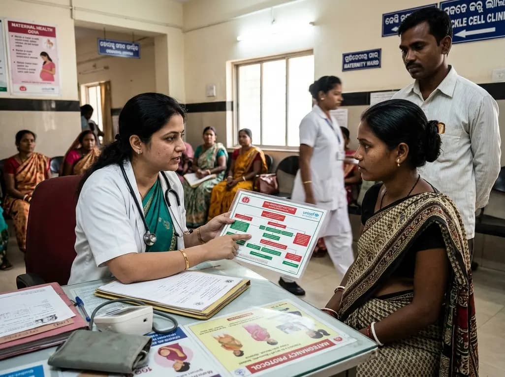
Odisha has established India's first state-level clinical protocol specifically tailored for pregnant women with sickle cell disease. Developed with NHM and UNICEF, the framework provides structured antenatal screening, crisis mitigation, standardized referral pathways, and specialized interdisciplinary care.
Today
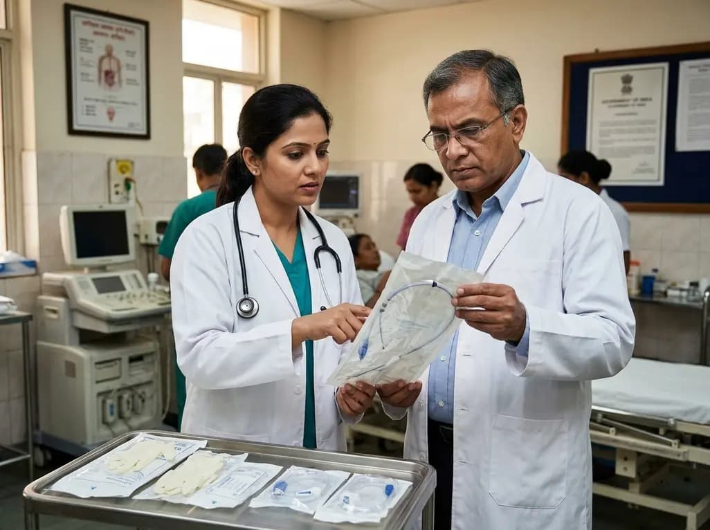
The Directorate General of Health Services directs Indian states to halt the hazardous reuse and reprocessing of single-use medical devices. Discover regulatory implications under the Drugs and Cosmetics Act, critical infection risks, legal liabilities, and operational changes required across clinical specialties.
Today

Mitochondria-targeted nanoparticles self-assembled from EGCG and L-glutamic acid effectively restore mitochondrial homeostasis, suppress synovial inflammation, and attenuate chondrocyte senescence in osteoarthritis, offering a promising disease-modifying nanotherapy.
Today

India is advancing preventive healthcare by integrating genomics, artificial intelligence, and quantum technologies. With the SANGAM programme sequencing one million Indian genomes, clinicians can unlock personalized therapies, detect cardiometabolic risks early, and curb the burden of diabetes and heart disease.
Today
