

Loading, please wait...
Loading, please wait...

A primary cauda equina neuroendocrine tumor represents an exceptionally rare intradural extramedullary neoplasm of the lumbosacral spine. Historically, pathologists and neurosurgeons classified these distinctive lesions as cauda equina paragangliomas. However, contemporary molecular insights and updated World Health Organization classifications have definitively categorized them as distinct neuroendocrine entities. Because their anatomical localization, clinical symptoms, and initial neuroimaging profiles closely mimic more frequent neoplasms, diagnostic confusion often arises. Consequently, clinicians must maintain a high index of suspicion when evaluating complex cauda equina syndromes.
Historically, medical literature referred to neuroendocrine lesions of the filum terminale as paragangliomas. However, the 2022 World Health Organization classification reclassified them as neuroendocrine neoplasms. This critical nomenclature transition reflects fundamental differences in cellular origin, genetic profile, and biologic behavior. Unlike traditional extra-adrenal sympathetic paragangliomas, these intradural tumors show diffuse immunoreactivity for cytokeratins such as CK8/18 and CAM5.2. Furthermore, they typically lack mutations in succinate dehydrogenase subunit genes, which frequently drive hereditary paraganglioma syndromes. Instead, these neoplasms frequently express HOXB13, indicating an origin related to embryological tail bud derivatives. In clinical practice, distinguishing primary cauda equina neuroendocrine tumors from true paragangliomas guides genetic screening strategies. While systemic paragangliomas mandate comprehensive germline testing for familial syndromes, primary lumbar neuroendocrine lesions typically arise as sporadic solitary masses. Therefore, clinicians avoid unnecessary hereditary panic when recognizing this distinct diagnostic entity. Neurosurgeons and pathologists must collaborate closely to ensure precise tumor categorization, preventing overtreatment while maintaining accurate longitudinal surveillance protocols.
Patients harboring these lumbar masses commonly present with progressive radiculopathy, chronic low back pain, and motor weakness. Frequently, nocturnal discomfort awakens patients from restorative sleep, serving as a subtle warning sign of spinal canal compromise. As tumor volume expands, compressive cauda equina syndrome eventually develops, manifesting as perineal numbness, bilateral leg heaviness, and urinary incontinence. Magnetic resonance imaging serves as the definitive initial diagnostic modality. On non-contrast sequences, the mass typically appears isointense on T1-weighted images and hyperintense on T2-weighted acquisitions. Moreover, post-contrast administration reveals intense, relatively uniform contrast enhancement due to extensive intrinsic vascularity. Importantly, prominent serpentine flow voids and dorsal eccentric vascular pedicles often surround the mass. However, these radiological features closely simulate myxopapillary ependymoma and spinal schwannoma. Both entities arise along the filum terminale and display robust contrast uptake. Consequently, radiologists and spine surgeons cannot rely solely on pre-operative imaging to establish a definitive tissue diagnosis. Recognizing prominent vascular engorgement nevertheless prepares the surgical team for substantial intraoperative bleeding.
Urgent microsurgical decompression represents the cornerstone of management when patients exhibit evolving neurological deficits. Surgeons typically perform a targeted laminoplasty or laminectomy across the involved vertebral levels. Intraoperatively, the tumor presents as an encapsulated, highly vascular, and friable mass adherent to the filum terminale and adjoining nerve roots. Therefore, microsurgical dissection demands meticulous technique, continuous electrophysiological nerve monitoring, and precise bipolar hemostasis. Because significant bleeding can obscure delicate neural planes, surgeons often identify and coagulate main feeding vessels early in the resection. Whenever feasible, gross-total resection remains the primary surgical objective, as complete excision confers excellent long-term disease-free survival. However, surgeons must avoid aggressive traction on compressed cauda equina roots to prevent permanent sphincter dysfunction and neurological deficit. If the lesion adheres densely to critical neural fibers, safe subtotal resection provides immediate neural decompression while limiting permanent postoperative morbidity. Postoperatively, patients frequently experience rapid functional recovery, with substantial improvement in lower extremity weakness and resolution of neuropathic radicular pain.
Definitive identification of these lesions depends entirely on rigorous microscopic examination and comprehensive immunohistochemical profiling. Histologically, the neoplasm demonstrates a classical organoid pattern composed of uniform cells with round nuclei, granular salt-and-pepper chromatin, and moderate eosinophilic cytoplasm. Reticulin staining highlights a nested architecture surrounded by capillary networks. Immunohistochemistry plays an indispensable role in separating this neoplasm from myxopapillary ependymoma, schwannoma, and metastatic lesions. The tumor cells show strong, diffuse positivity for neuroendocrine markers, including synaptophysin and chromogranin A. Additionally, cytokeratin markers CK8/18 and CAM5.2 reveal characteristic dot-like or perinuclear positivity. In contrast, glial fibrillary acidic protein remains consistently negative, effectively excluding ependymoma. Furthermore, S100 and SOX10 stains highlight delicate sustentacular cells around tumor nests rather than the neoplastic cells themselves, distinguishing the lesion from schwannoma. Pathologists also evaluate the Ki-67 proliferation index, which typically remains under two percent in WHO grade 1 neoplasms. Therefore, this targeted panel establishes the diagnosis with outstanding specificity and reproducibility.
Following histopathological confirmation, clinicians must immediately rule out metastatic neuroendocrine disease. While primary lumbar neuroendocrine lesions behave indolently, spinal drop metastases or hematogenous deposits from gastroenteropancreatic neuroendocrine tumors can mimic primary lesions. Therefore, patients should undergo comprehensive postoperative staging, including contrast-enhanced computed tomography of the chest, abdomen, and pelvis. Additionally, gallium-68 DOTATATE positron emission tomography provides extraordinary sensitivity for identifying occult primary sites or distant systemic metastases. When postoperative staging confirms an isolated lumbar lesion with clear surgical margins, clinicians rarely recommend adjuvant radiation therapy. Instead, clinicians institute structured longitudinal imaging protocols using contrast-enhanced spinal magnetic resonance imaging. Although recurrence after gross-total excision remains exceedingly rare, late recurrences and delayed drop metastases have appeared several years post-resection. Consequently, experts recommend extended clinical and radiological follow-up for at least five to ten years. Multidisciplinary collaboration among spine surgeons, oncologists, and endocrinologists ensures comprehensive, individualized long-term care for every affected individual.
Most primary cauda equina neuroendocrine tumors are functionally non-secretory, so patients rarely present with systemic endocrine syndromes. However, rare functioning tumors can release vasoactive catecholamines, serotonin, or neuropeptides. In such uncommon instances, individuals may experience episodic hypertension, palpitations, severe headaches, or facial flushing. Therefore, clinicians should obtain preoperative fractionated plasma metanephrines if a patient displays labile blood pressure or unexplained tachycardia during initial clinical assessment.
Clinically and radiologically, both neoplasms share overlapping presentations, including nocturnal back pain, radiculopathy, and well-circumscribed lumbar intradural enhancement. However, myxopapillary ependymomas typically exhibit a slightly higher propensity for cerebrospinal fluid dissemination and local recurrence, carrying a WHO grade 2 classification. Conversely, primary neuroendocrine tumors carry a WHO grade 1 designation, demonstrating negative glial fibrillary acidic protein staining and distinct cytokeratin positivity that conclusively distinguish them pathologically.
The long-term prognosis following gross-total resection of a primary neuroendocrine tumor is exceptionally favorable. Because these lesions are low-grade neoplasms with minimal proliferative activity, complete surgical excision typically provides a definitive cure without requiring adjuvant radiotherapy or systemic chemotherapy. Nevertheless, clinicians recommend periodic magnetic resonance surveillance for at least five to ten years, as late local recurrences and rare subarachnoid seedings have occurred.
Disclaimer: This content is for informational and educational purposes only. It is not intended to be a substitute for professional medical advice, diagnosis, or treatment. Refer to the latest local and national guidelines for clinical practice.
References

Read summarized clinical updates, watch expert medical content, and earn CME certifications right from your smartphone.
Open your smartphone camera to scan and install.





Primary cauda equina neuroendocrine tumors (CENETs) are rare intradural neoplasms that frequently mimic myxopapillary ependymomas. Complete surgical excision, detailed immunohistochemistry, and systemic staging are crucial to ensure optimal patient outcomes and distinguish them from metastatic disease.
Today

Despite proven efficacy in preventing severe morbidity and mortality, adult immunization coverage among Indian seniors remains below two percent. Pulmonologists and geriatricians urge widespread adoption of seasonal influenza, pneumococcal conjugate, and recombinant zoster vaccines to curb preventable hospitalizations.
Today

An intracranial EWSR1::CREM fusion sarcoma in a 31-year-old female manifested as a paraneoplastic inflammatory syndrome with anemia and secondary infertility. The dural mass mimicked meningioma, but gross-total resection resolved her systemic inflammation and restored natural fertility within one month.
Today

The DOMINO study protocol presents a multidisciplinary implementation mapping approach to overcome provider, patient, and payer barriers to antiobesity medications, offering vital insights for Indian clinicians managing the growing diabesity burden.
Today

A comprehensive analysis of MAP therapy resistance in osteosarcoma, examining canonical and non-canonical mechanisms across damage avoidance, enhanced DNA repair, and replication stress tolerance to identify novel vulnerabilities.
Today
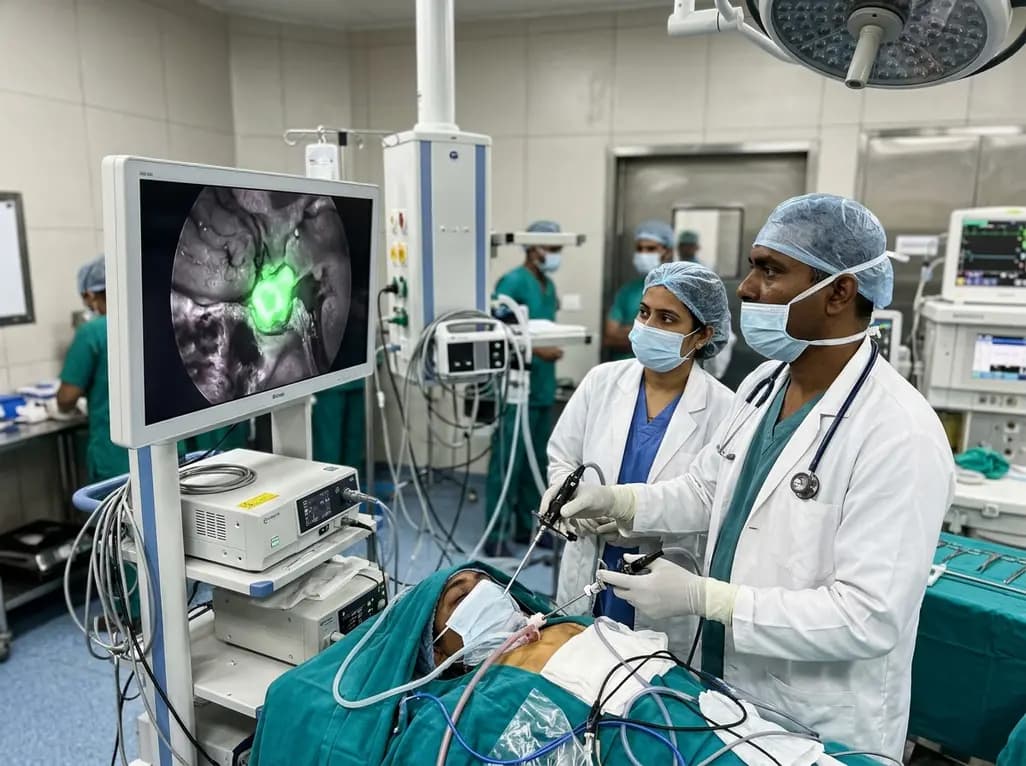
Near-infrared autofluorescence provides real-time label-free optical guidance during endoscopic transsphenoidal pituitary surgery. By highlighting stark signal contrasts between normal pituitary gland and adenomas, this technique aids gross-total tumor resection while safeguarding critical endocrine function.
Today
