

Loading, please wait...
Loading, please wait...

Flow-diverter stents have revolutionized endovascular management for complex intracranial aneurysms. However, neurointerventional teams must remain vigilant regarding unexpected flow-diverter stent complications. While flow diverters primarily induce hemodynamic stagnation to promote intra-aneurysmal thrombosis, downstream or parent vessel events can still emerge. Most literature focuses on small branch vessel occlusions, which seldom cause permanent neurological deficits. In contrast, late thrombotic occlusion of proximal large vessels represents a devastating clinical scenario. A recently published clinical case details an unusual presentation where a patient developed delayed common carotid artery occlusion one year after device implantation. Understanding the etiology, clinical manifestations, and preventive strategies is essential for clinicians who manage neurovascular disease.
Endovascular specialists frequently utilize fine-mesh devices like the Surpass Evolve system to manage wide-necked aneurysms. Consequently, these devices reconstruct the parent vessel by reducing intra-aneurysmal shear stress. Over time, endothelial cells proliferate across the stent struts, thereby isolating the aneurysm from arterial circulation. Nevertheless, flow-diverter stent complications can arise when device-induced flow dynamics trigger unintended thrombogenesis. In typical anterior circulation cases, covered branch arteries may undergo gradual remodeling without causing overt ischemic deficits. In contrast, common carotid artery occlusion following distal intracranial stenting remains an exceedingly rare phenomenon. Several factors might precipitate this proximal occlusion. For example, local endothelial injury during catheter manipulation can generate an unstable thrombogenic substrate. Additionally, microemboli or retrograde propagation of thrombus may compromise upstream arterial flow. Clinicians must recognize that stent porosity and braid design influence vascular biology. Therefore, understanding device-tissue interactions is vital to prevent catastrophic vascular events.
In the highlighted case report, the patient presented one year after device deployment with acute motor impairment. Specifically, the individual experienced unilateral limb weakness, prompting urgent medical evaluation. Cranial magnetic resonance imaging subsequently revealed a left frontoparietal cerebral infarction. Furthermore, neurovascular imaging demonstrated complete occlusion of the ipsilateral common carotid artery. This delayed presentation surprised the clinical team because thrombotic complications usually cluster within the early postoperative period. Therefore, physicians must maintain a high index of suspicion even years after successful aneurysm exclusion. Symptoms often mimic routine thromboembolic strokes, manifesting as focal motor deficits, aphasia, or altered sensory perception. Moreover, collateral circulation significantly determines the severity of clinical manifestations. Patients with robust circle of Willis architecture may remain asymptomatic, whereas those with poor collateral flow suffer major tissue hypoxia. Hence, regular clinical assessments remain imperative throughout extended follow-up periods.
Researchers continue to investigate the precise mechanisms driving proximal vessel thrombosis after distal flow diversion. First, delayed in-stent thrombosis may propagate retrogradely along the internal carotid axis into the common carotid artery. Second, altered hemodynamics and turbulent flow patterns around the device may stimulate persistent platelet aggregation. Furthermore, patient-specific systemic factors heavily influence late thrombotic risks. For instance, hyporesponsiveness to antiplatelet therapy significantly increases vascular vulnerability. Atherosclerotic plaques located near the carotid bifurcation can also rupture under altered shear stress environments. Consequently, platelet deposition accelerates, leading to complete arterial occlusion. In addition, intimal hyperplasia and delayed endothelization can foster chronic thromboembolic foci. Therefore, clinicians must evaluate both anatomical geometry and systemic coagulation tendencies. When structural stenosis coexists with hypercoagulability, the risk of extensive carotid thrombosis escalates dramatically.
Accurate neurovascular imaging forms the foundation of post-procedure surveillance following flow diversion. Initially, magnetic resonance angiography and computed tomography angiography provide detailed anatomical assessments of stent patency. However, digital subtraction angiography remains the reference standard for evaluating subtle hemodynamic alterations and collateral reconstitution. Furthermore, transcranial Doppler ultrasonography offers non-invasive monitoring of intracranial flow velocities. Clinicians should establish structured follow-up timelines, typically scheduling evaluations at three, six, and twelve months post-implantation. If patients exhibit new neurological deficits, emergency magnetic resonance imaging with diffusion-weighted sequences must occur immediately. Additionally, perfusion imaging helps determine salvageable brain tissue within ischemic penumbras. Because late arterial occlusions can develop silently, routine vascular imaging detects evolving parent vessel stenosis before clinical infarction occurs. Consequently, strict adherence to surveillance protocols enables timely therapeutic interventions.
Pharmacotherapy serves as the cornerstone of stroke prevention after endovascular flow diversion. Typically, clinicians prescribe dual antiplatelet therapy consisting of aspirin and a P2Y12 inhibitor prior to stent placement. This combination prevents acute platelet aggregation on metallic mesh surfaces. Subsequently, practitioners taper therapy to single antiplatelet prophylaxis once adequate endothelial coverage develops. In the reported case, the patient received enteric-coated aspirin tablets following the diagnosis of cerebral infarction. Fortunately, the individual demonstrated notable symptomatic improvement and achieved safe hospital discharge. Nonetheless, the optimal duration of prolonged antiplatelet therapy remains an ongoing debate among neurointerventionalists. Moreover, platelet function testing assists in identifying resistant non-responders who need alternative antiplatelet agents such as ticagrelor or prasugrel. Therefore, personalized antithrombotic regimens are essential to prevent late ischemic strokes while mitigating intracranial hemorrhage risks.
Carotid artery occlusion after flow-diverter stent placement is an extremely rare complication. Most flow diversion complications involve acute in-stent thrombosis or small covered branch occlusions, which seldom cause permanent neurological injury. Late occlusion of the proximal common carotid artery occurs in only isolated clinical reports. Clinicians must nevertheless remain vigilant, as sudden proximal occlusion can cause devastating cerebral infarction if collateral cerebral circulation proves inadequate.
Delayed arterial occlusion typically manifests as acute ischemic stroke symptoms. Patients frequently develop sudden focal neurological deficits, including unilateral limb weakness, facial droop, numbness, or impaired coordination. In addition, patients may experience expressive or receptive aphasia, visual field disturbances, and acute dizziness. Any new or progressive neurological impairment occurring months or years after endovascular treatment demands urgent emergency neurovascular evaluation to prevent permanent cerebral injury.
Standard protocol mandates dual antiplatelet therapy with aspirin and clopidogrel or ticagrelor before and immediately following stent implantation. Interventional teams maintain this dual regimen for three to six months to prevent acute thrombogenesis while endothelialization occurs. Afterward, clinicians generally transition patients to lifelong single antiplatelet therapy. In addition, platelet function testing helps identify drug resistance, allowing personalized dosing regimens that reduce both ischemic and hemorrhagic complications.
Disclaimer: This content is for informational and educational purposes only and should not be considered as medical advice or a substitute for professional healthcare guidance. Always consult with a qualified physician or healthcare provider for diagnosis and treatment options suited to your individual needs. The information provided here is based on available data and research at the time of publication, but medicine is an ever-evolving field, and standards of care may change. While every effort has been made to ensure accuracy, neither the authors nor the publishers assume any liability for any harm, injury, or adverse effects arising from the application or interpretation of the material herein. Refer to the latest local and national guidelines for clinical practice.
References

Read summarized clinical updates, watch expert medical content, and earn CME certifications right from your smartphone.
Open your smartphone camera to scan and install.





A rare case report highlights common carotid artery occlusion and cerebral infarction occurring one year after flow-diverter stent placement for an intracranial aneurysm. Prompt medical management restored neurological function, underscoring the vital need for vigilant vascular surveillance.
Today

Despite proven efficacy in preventing severe morbidity and mortality, adult immunization coverage among Indian seniors remains below two percent. Pulmonologists and geriatricians urge widespread adoption of seasonal influenza, pneumococcal conjugate, and recombinant zoster vaccines to curb preventable hospitalizations.
Today

An intracranial EWSR1::CREM fusion sarcoma in a 31-year-old female manifested as a paraneoplastic inflammatory syndrome with anemia and secondary infertility. The dural mass mimicked meningioma, but gross-total resection resolved her systemic inflammation and restored natural fertility within one month.
Today

The DOMINO study protocol presents a multidisciplinary implementation mapping approach to overcome provider, patient, and payer barriers to antiobesity medications, offering vital insights for Indian clinicians managing the growing diabesity burden.
Today

A comprehensive analysis of MAP therapy resistance in osteosarcoma, examining canonical and non-canonical mechanisms across damage avoidance, enhanced DNA repair, and replication stress tolerance to identify novel vulnerabilities.
Today
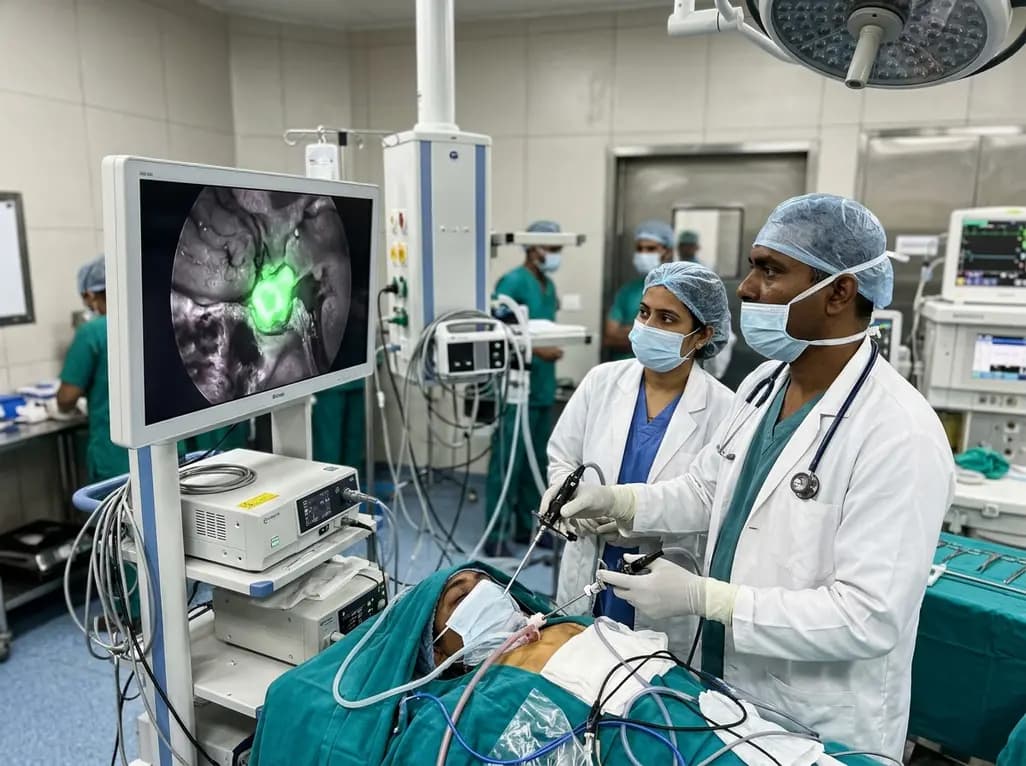
Near-infrared autofluorescence provides real-time label-free optical guidance during endoscopic transsphenoidal pituitary surgery. By highlighting stark signal contrasts between normal pituitary gland and adenomas, this technique aids gross-total tumor resection while safeguarding critical endocrine function.
Today
