

Loading, please wait...
Loading, please wait...

Managing acute ischemic stroke secondary to cardioembolism presents an urgent clinical dilemma. Clinicians must constantly balance early thrombus propagation against catastrophic hemorrhagic transformation. Therefore, accurately estimating cardioembolic stroke recurrence remains essential during initial hospital admission. While neurological impairment offers immediate diagnostic bedside insight, neuroimaging provides objective structural details about cerebral tissue destruction. Historically, physicians relied heavily on the National Institutes of Health Stroke Scale (NIHSS) to gauge baseline disease severity. However, functional impairment on admission does not always mirror the exact anatomical extent of ischemic parenchymal damage. For instance, small subcortical lesions in eloquent motor pathways can produce profound clinical deficits. Conversely, substantial territorial infarctions within non-dominant parietal lobes may manifest with deceptively moderate neurological signs. Consequently, determining whether anatomical lesion size or clinical deficit better predicts secondary events has generated substantial clinical interest. A multicenter cohort investigation encompassing 2,824 acute cardioembolic stroke patients evaluated this critical prognostic question. By examining both clinical scores and radiologic dimensions, researchers established valuable prognostic benchmarks for contemporary inpatient stroke units. Understanding these nuances helps clinicians anticipate secondary deterioration and personalize acute therapeutic pathways effectively.
To dissect these prognostic dynamics, investigators conducted a comprehensive observational study analyzing 2,824 individuals presenting with acute cardioembolic stroke. Researchers stratified neurological severity into three distinct cohorts based on admission NIHSS scores: minor stroke (NIHSS 0 to 4), moderate stroke (NIHSS 5 to 15), and severe stroke (NIHSS greater than 15). Simultaneously, clinicians categorized baseline neuroimaging infarct size into small, medium, or large volume thresholds. The primary outcome was a composite 90-day clinical endpoint encompassing either ischemic complications or hemorrhagic events. Specifically, ischemic endpoints captured recurrent ischemic stroke and systemic embolism, whereas hemorrhagic endpoints captured symptomatic intracranial hemorrhage or severe extracranial bleeding. Overall, the investigators recorded 230 composite primary outcome events during the 90-day follow-up period, representing an overall event incidence of 8.1 percent. Moreover, this substantial event frequency underscores the persistent vulnerability of cardioembolic patients during the early convalescent phase. Because post-stroke morbidity spikes drastically after secondary vascular events, robust baseline risk stratification proves crucial. Consequently, comparing the distinct predictive contributions of radiologic and clinical markers illuminates how clinicians can better identify high-risk individuals requiring vigilant early hemodynamic and neurological observation.
Multivariable regression models revealed clear associations between baseline characteristics and 90-day adverse outcomes. Specifically, with the minor stroke category (NIHSS 0 to 4) serving as the reference group, moderate (NIHSS 5 to 15) and severe deficits (NIHSS greater than 15) independently predicted both composite and hemorrhagic outcomes. Furthermore, the severe stroke category correlated strongly with recurrent ischemic events. Meanwhile, when small radiographic lesions served as the baseline reference, multivariable analysis demonstrated that medium and large lesions correlated significantly with all evaluated outcomes, including ischemic, hemorrhagic, and composite endpoints. Receiver operating characteristic (ROC) analyses offered deeper comparative insights into prognostic precision. For the primary composite outcome, infarct size achieved an area under the curve (AUC) of 0.618, whereas admission NIHSS yielded an AUC of 0.599. Similarly, for hemorrhagic complications, lesion size demonstrated superior discrimination with an AUC of 0.649 compared to 0.601 for NIHSS. Although DeLong testing indicated that these AUC discrepancies did not reach formal statistical significance, radiologic lesion size numerically outperformed neurological scoring. For purely ischemic outcomes, both parameters demonstrated comparable, modest predictive discrimination, yielding AUC values of 0.575 and 0.577, respectively.
The observed data highlight a fundamental therapeutic tension in early stroke care. Patients who present with extensive parenchymal damage harbor damaged microvasculature, leaky capillary beds, and compromised blood-brain barrier integrity. Consequently, this pathophysiologic vulnerability markedly amplifies the threat of petechial extravasation and secondary symptomatic hematoma formation. However, clinicians cannot overlook systemic thrombotic hazards, because underlying atrial fibrillation continually threatens recurrent cerebral embolization. Current international stroke guidance frequently promotes clinical scoring tools to govern early oral anticoagulation initiation. Nevertheless, these newer findings emphasize that relying solely on bedside neurological exams can misestimate bleeding vulnerability. For example, a patient with mild speech hesitation may nevertheless possess a large silent cortical infarction susceptible to severe hemorrhagic transformation upon premature anticoagulation. Conversely, extensive visual neglect might stem from localized vascular occlusion that entails lower hemorrhagic hazards than global territorial necrosis. Therefore, integrating routine volumetric brain imaging into therapeutic algorithms provides vital safety margins. Clinicians must weigh the verified anatomical destruction before prescribing targeted direct oral anticoagulants or therapeutic heparins. Balancing these dual risks prevents tragic iatrogenic bleeding while maintaining effective secondary stroke prevention.
These findings hold profound practical implications for acute stroke care throughout India. In Indian healthcare settings, non-valvular atrial fibrillation and rheumatic heart disease represent prominent drivers of cardioembolic strokes. Additionally, patients frequently present with delayed hospital arrival times, which restricts acute reperfusion opportunities and expands final infarct dimensions. Because public and private emergency centers across urban and semi-urban India increasingly maintain twenty-four-hour computed tomography and magnetic resonance imaging facilities, radiologic lesion assessment is highly feasible. However, clinicians often face challenging decisions regarding anticoagulant initiation due to variable patient compliance, monitoring limitations, and diverse socioeconomic barriers. Relying exclusively on admission NIHSS may lead doctors to initiate anticoagulation prematurely in patients exhibiting mild clinical deficits despite harboring substantial infarct sizes. Furthermore, unmonitored anticoagulation in large infarcts carries high fatality rates from hemorrhagic conversion. Therefore, multidisciplinary Indian stroke teams should prioritize standardized neuroimaging re-evaluation before starting anticoagulants. Adopting repeat non-contrast CT or diffusion-weighted MRI scans allows clinicians to verify structural stability and exclude silent hemorrhagic conversion. Such structured diagnostic protocols optimize clinical outcomes, particularly within resource-constrained secondary and tertiary care centers.
To translate these scientific findings into routine hospital practice, clinicians should adopt a comprehensive, multidisciplinary management approach. First, physicians must systematically document both admission NIHSS scores and anatomical lesion volumes during initial emergency evaluations. Second, stroke teams should avoid relying solely on clinical severity scores when planning the timing of oral anticoagulation. Instead, clinicians should incorporate imaging-based classifications, categorizing lesions into small, medium, and large volumes to titrate antithrombotic timing safely. Third, clinicians ought to perform follow-up neuroimaging around days three to seven for moderate-to-large infarctions prior to commencing anticoagulant therapy. This precaution reliably rules out occult petechial bleeding or substantial edema before antithrombotic exposure. Fourth, treating physicians must optimize comorbid blood pressure, glycemic parameters, and hydration, because microvascular stress accelerates both infarct expansion and secondary extravasation. Furthermore, continuous cardiac telemetry remains mandatory to detect paroxysmal atrial fibrillation in cryptogenic embolic strokes. Finally, clear communication with patients and families regarding early warning signs of recurrent ischemia and bleeding enhances long-term compliance. By combining refined radiological metrics with structured neurological assessments, clinicians can significantly mitigate adverse outcomes across acute cardioembolic stroke populations.
In clinical trials, radiologic lesion size slightly outperformed admission NIHSS scores in predicting 90-day composite and hemorrhagic complications. Although statistical significance was not reached on DeLong testing, infarct volume provides crucial structural information about blood-brain barrier integrity that bedside clinical examinations alone cannot fully capture.
Anticoagulation timing depends heavily on infarct size and bleeding risk. While minor infarctions often permit initiation within three to four days, medium and large territorial lesions require delayed anticoagulation, typically after seven to fourteen days. Clinicians should confirm absence of hemorrhagic transformation using repeat neuroimaging prior to therapy.
Severe cardioembolic stroke reflects substantial underlying thrombus burden and extensive cerebral tissue necrosis. Extensive infarction severely disrupts endothelial microvasculature, heightening susceptibility to reperfusion bleeding. Concurrently, the persistent cardiac thrombogenic substrate and pronounced systemic inflammatory responses drive further thromboembolism, amplifying recurrent ischemic risks during the acute 90-day period.
Disclaimer: This content is for informational and educational purposes only and should not be considered medical advice. Healthcare professionals should exercise their independent clinical judgment. Refer to the latest local and national guidelines for clinical practice.
References

Read summarized clinical updates, watch expert medical content, and earn CME certifications right from your smartphone.
Open your smartphone camera to scan and install.





In a cohort of 2,824 cardioembolic stroke patients, infarct lesion size predicted 90-day recurrence and bleeding slightly better than admission NIHSS score. Clinicians must integrate neuroimaging with neurological exams to balance ischemic recurrence prevention against catastrophic hemorrhagic transformation.
Today
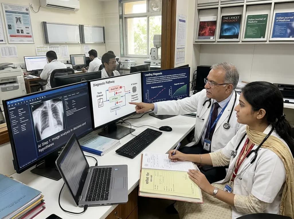
A breakthrough peer-reviewed study demonstrates that a sequential AI diagnosis system can mirror real-world clinical reasoning. By ordering stepwise tests and updating hypotheses dynamically, this multi-agent orchestrator achieved superior diagnostic accuracy while reducing investigative costs.
Today

A prenatal case highlights severe glutaric acidemia type II presenting as bilateral hyperechoic cystic kidneys at 18 weeks. Whole-exome sequencing revealed a novel homozygous ETFDH frameshift variant, demonstrating the power of rapid genetic testing in fetal renal anomalies.
Today

Review how tailored exercise programs benefit frail older patients with heart failure. Explore pathophysiological mechanisms, individualized training prescriptions, and strategies to overcome adherence barriers in clinical practice.
Yesterday

This comparative study evaluates posterior labral tears with and without glenohumeral instability, highlighting distinct demographic patterns, MRI underestimation in stable shoulders, and successful arthroscopic repair outcomes.
Yesterday

A clinical study demonstrates that early intervention with tocilizumab, either as monotherapy or bridged with corticosteroids, achieves significant proptosis reduction and resolves active inflammation in treatment-naive thyroid eye disease.
Yesterday
