.webp&w=384&q=75)

Loading, please wait...
Loading, please wait...
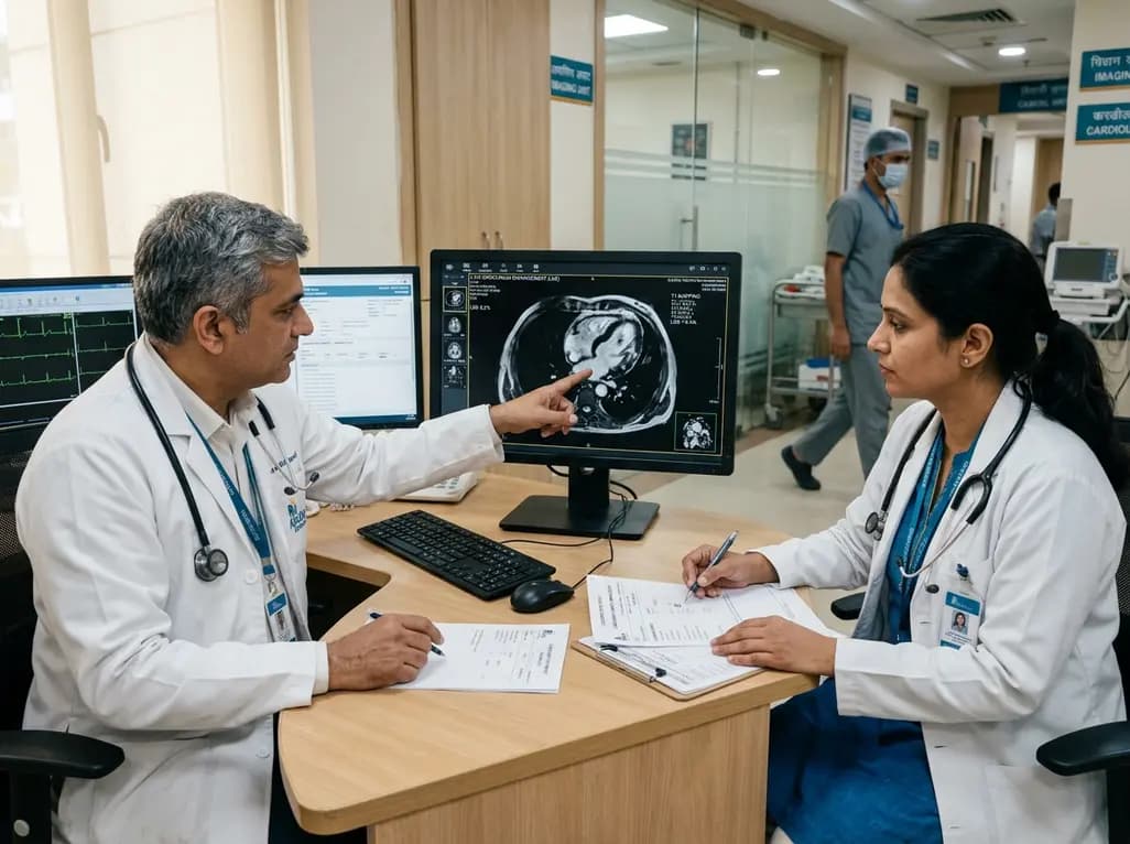
Acute myocarditis presents clinicians with significant diagnostic and prognostic hurdles due to its diverse clinical spectrum, ranging from mild chest discomfort to life-threatening cardiogenic shock. Clinicians frequently utilize non-invasive cardiovascular imaging to evaluate myocardial inflammation and necrosis. Advanced cardiac MRI acute myocarditis protocols now provide unparalleled tissue characterization, helping clinicians distinguish between reversible inflammatory edema and permanent fibrotic injury. When clinicians combine quantitative tissue parameters with circulating inflammatory biomarkers like C-reactive protein, they achieve superior risk stratification. This approach enables timely interventions, optimizes the deployment of mechanical circulatory support, and accurately predicts adverse long-term cardiovascular outcomes.
Cardiovascular magnetic resonance imaging serves as the non-invasive gold standard for assessing myocardial inflammation and structural damage. Clinicians rely on multiparametric protocols, including cine imaging, native T1 mapping, T2 mapping, post-contrast T1 mapping, and late gadolinium enhancement, to assess cardiac integrity. Native T1 relaxation times and extracellular volume fractions quantify diffuse interstitial expansion and microscopic collagen deposition. Simultaneously, T2 mapping precisely detects myocardial edema, which reflects the active inflammatory phase of the disease. In contrast, late gadolinium enhancement highlights focal areas of regional necrosis and replacement fibrosis.
Furthermore, cardiac imaging differentiates active inflammatory changes from irreversible myocardial damage. Although myocardial edema often resolves over several weeks, persistent replacement fibrosis indicates established structural injury. Consequently, comprehensive tissue mapping helps clinicians track disease resolution and guide therapeutic intensity. By providing quantitative assessments of extracellular volume and tissue relaxation times, advanced magnetic resonance protocols offer profound physiological insight into the myocardial microenvironment. Therefore, standardizing these advanced imaging sequences within tertiary referral centers significantly improves diagnostic accuracy and facilitates early risk assessment.
The clinical trajectory of acute myocarditis varies substantially depending on whether the disease takes a fulminant or non-fulminant course. Fulminant myocarditis presents abruptly with severe hemodynamic collapse, severe left ventricular dysfunction, and intractable arrhythmias, frequently demanding prompt mechanical circulatory support. In contrast, non-fulminant myocarditis follows an indolent course with milder symptoms and lower immediate mortality.
Recent clinical investigations demonstrate that patients who present with fulminant myocarditis requiring mechanical circulatory support exhibit markedly elevated native T1, extracellular volume fractions, and late gadolinium enhancement percentages compared to non-fulminant cases. Interestingly, quantitative T2 values representing myocardial edema remain comparable across both clinical groups. This crucial finding indicates that fulminant presentations driven by cardiogenic shock do not simply involve greater tissue water content. Instead, these severe cases carry a substantially higher burden of cellular necrosis and acute interstitial matrix disruption. Consequently, clinicians must recognize that the absolute extent of fibrotic scar and necrosis drives hemodynamic instability rather than inflammatory edema alone.
The quantitative extent of late gadolinium enhancement provides clinicians with robust prognostic data regarding long-term cardiovascular events. Studies demonstrate that a threshold of late gadolinium enhancement exceeding 8.4 percent of left ventricular mass independently predicts major adverse cardiovascular events and functional decline. Patients surpassing this cut-off face heightened risks of sustained ventricular arrhythmias, heart failure hospitalizations, and long-term functional impairment classified as New York Heart Association functional classes II to IV.
Moreover, incorporating quantitative enhancement percentages into multivariate prognostic models significantly enhances predictive accuracy, elevating the C-statistic from baseline levels of 0.74 up to 0.82. This incremental predictive power underscores the clinical utility of measuring exact scar burden rather than relying merely on visual evaluations. Focal necrosis acts as a durable substrate for re-entrant ventricular tachyarrhythmias and progressive left ventricular remodeling. Consequently, clinicians should obtain late gadolinium enhancement quantification early during the convalescent phase to guide rhythm surveillance. Therefore, cardiac magnetic resonance quantification provides an indispensable tool for tailoring individualized follow-up intervals.
Circulating inflammatory biomarkers provide critical complementary information to anatomical and tissue imaging. Serum C-reactive protein serves as an accessible systemic marker that reflects acute vascular and systemic inflammatory cascades. When clinicians identify highly elevated baseline C-reactive protein levels, specifically exceeding 67.9 mg/dL, in conjunction with significant myocardial scar burden, they can identify patients at exceptionally high risk for adverse outcomes.
Furthermore, integrating systemic inflammation with tissue-level fibrotic metrics creates a multidimensional risk stratification framework. While elevated C-reactive protein highlights active systemic immune responses, myocardial enhancement reflects localized structural destruction. Patients exhibiting both high inflammatory activity and extensive tissue necrosis experience the highest incidence of subsequent heart failure. Conversely, patients with low systemic inflammatory markers and minimal enhancement demonstrate favorable functional recovery. Therefore, combining routine laboratory assays with cardiac magnetic resonance parameters improves prognostic accuracy without imposing unreasonable diagnostic burdens on healthcare systems.
Integrating advanced cardiac imaging and inflammatory biomarker screening into standard clinical workflows substantially enhances inpatient and outpatient management. During the acute admission, identifying severe tissue expansion and substantial scar burden helps intensive care specialists determine the optimal timing for invasive hemodynamic monitoring or mechanical circulatory assistance. Furthermore, clinicians can utilize these baseline parameters to identify patients who may benefit from targeted anti-inflammatory regimens or aggressive neurohormonal blockade.
Following hospital discharge, risk-stratified patients require tailored longitudinal care to prevent sudden cardiac death and chronic inflammatory cardiomyopathy. Individuals with late gadolinium enhancement exceeding the critical prognostic threshold should undergo extended ambulatory rhythm monitoring and scheduled echocardiographic reassessments. In addition, clinicians must advise strict exercise restriction during the acute phase until repeat imaging confirms the resolution of active inflammation. Consequently, multiparametric cardiovascular magnetic resonance combined with serial biomarker tracking enables personalized clinical pathways.
Late gadolinium enhancement reflects localized myocardial necrosis and irreversible replacement fibrosis. A high percentage of enhancement, particularly exceeding 8.4 percent of left ventricular mass, independently correlates with major adverse cardiovascular events, recurrent hospitalizations, and persistent functional decline. Consequently, quantifying enhancement helps clinicians identify high-risk individuals who require vigilant arrhythmia surveillance and structured medical therapy to prevent long-term post-inflammatory heart failure complications.
Serum C-reactive protein measures systemic inflammatory activity during the acute disease phase, whereas magnetic resonance imaging captures localized tissue destruction and scar burden. When clinicians combine elevated C-reactive protein levels above 67.9 mg/dL with substantial myocardial late gadolinium enhancement, they gain superior prognostic accuracy. This synergistic combination improves clinical risk stratification, enabling medical teams to optimize patient monitoring and timely intensive care interventions.
Fulminant myocarditis requiring mechanical support exhibits significantly higher native T1 relaxation times, increased extracellular volume fractions, and greater late gadolinium enhancement compared to non-fulminant cases. Interestingly, both conditions show comparable levels of myocardial edema on T2 mapping. This demonstrates that fulminant disease severity is primarily driven by extensive cell necrosis and profound interstitial collagen disruption rather than the degree of tissue water accumulation alone.
Disclaimer: This content is for informational and educational purposes only and is intended exclusively for healthcare professionals. It should not be used as a substitute for professional medical judgment, diagnosis, or treatment. Refer to the latest local and national guidelines for clinical practice.
References

Read summarized clinical updates, watch expert medical content, and earn CME certifications right from your smartphone.
Open your smartphone camera to scan and install.





Cardiovascular magnetic resonance tissue mapping combined with C-reactive protein improves risk stratification in acute myocarditis. Late gadolinium enhancement above 8.4% and elevated CRP independently predict major adverse cardiovascular events and functional decline in adult patients.
last month

Pancreatic cancer cachexia affects over 80% of patients, driving severe muscle wasting, systemic inflammation, and treatment intolerance. This review highlights key pathophysiological drivers, exocrine insufficiency, objective CT imaging, and emerging therapies like GDF-15 inhibitors to improve clinical outcomes.
Today

A scoping review reveals how pregnancy affects phonation through laryngeal edema and reduced phonation time rather than major acoustic pitch alterations. Learn how to identify, evaluate, and manage vocal symptoms in expectant mothers.
Today

According to WHO data, average lifespan reached 73.3 years in 2023, recovering from pandemic lows. However, healthy life expectancy lags at 62.8 years due to surging noncommunicable conditions like cardiovascular disease and dementia, demanding proactive primary screening and sustained chronic care.
Today

Researchers have engineered an artificial sensory-pain integrated receptor using a multi-threshold organic synaptic transistor. The system distinguishes benign touch from noxious stimuli, enabling safer, more responsive prosthetic limbs with built-in memory and simplified hardware architecture.
Today

Medical robotics is transitioning beyond operative suites into diagnostics, cardiology, and endocrinology. Explore how innovations in tele-robotics, AI integration, and nanomedicine are overcoming geographical barriers and transforming precision healthcare delivery across India's evolving disease landscape.
Today
