.webp&w=384&q=75)

Loading, please wait...
Loading, please wait...

Surgical resection and stabilization of primary spinal neoplasms present complex reconstructive dilemmas for spine surgeons and oncologists. Recently, carbon fiber spinal instrumentation, composed of carbon fiber-reinforced polyetheretherketone (CFR-PEEK), has emerged as a compelling alternative to conventional titanium constructs. While titanium implants have long served as the benchmark for spinal stabilization, their metallic properties generate substantial diagnostic artifacts on magnetic resonance imaging (MRI) and computed tomography (CT). Furthermore, titanium causes considerable radiation beam scattering during adjuvant radiotherapy. Consequently, clinicians are increasingly adopting radiolucent carbon fiber implants to optimize postoperative oncologic surveillance and improve precision radiotherapy delivery.
Surgical management of primary spinal tumors demands stable biomechanical reconstruction following en bloc or intralesional tumor resection. Traditional metallic systems provide reliable structural support, yet they significantly interfere with post-surgical management. In particular, heavy metals produce dense scatter artifacts on cross-sectional imaging, which obscuring critical anatomical zones adjacent to the treated margins. Consequently, detecting early local recurrence becomes difficult for radiologists and oncologists. In addition, when patients require adjuvant radiation therapy, metallic pedicle screws induce perturbation and attenuation of ionizing radiation beams, leading to potential underdosing of tumor beds and overdosing of adjacent neural structures. Carbon fiber spinal instrumentation directly addresses these technological hurdles. Because carbon fiber-reinforced polymers exhibit radiolucency and low magnetic susceptibility, they produce minimal imaging artifacts. Moreover, their physical density allows therapeutic radiation beams to pass cleanly with negligible scattering. Despite these distinct imaging and dosimetric benefits, surgeons have sought definitive long-term clinical data to confirm that CFR-PEEK hardware achieves equivalent mechanical safety and tumor control relative to time-tested titanium constructs.
To evaluate the comparative performance of carbon fiber constructs in real-world clinical practice, researchers analyzed prospective multicenter data from the AO Spine Primary Tumor Research and Outcomes Network (PTRON). The investigator group identified a robust international cohort of 359 consecutive patients undergoing surgery for primary spinal neoplasms who met all inclusion criteria. Among the participants, 84 patients (23%) received carbon fiber spinal implants, 243 patients (68%) received conventional titanium implants, and 32 patients (9%) received hybrid instrumentation combining both materials. The median clinical follow-up reached 1.9 years in the carbon fiber cohort, 1.3 years in the titanium cohort, and 1.1 years in the combination group. The primary study endpoint focused on the overall incidence of postoperative adverse events. Additionally, secondary endpoints assessed the specific risk of sustaining at least one complication, local tumor recurrence, progression-free survival, and overall survival. Multivariable Cox proportional hazards regression models and Kaplan-Meier survival analyses were employed to control for potential confounding variables across clinical centers.
A primary concern among spine specialists adopting CFR-PEEK implants has been hardware failure, because early-generation composite devices raised theoretical questions about screw pullout and rod fatigue. However, the AO Spine PTRON analysis provides reassuring evidence regarding structural safety. Specifically, the estimated risk of developing at least one postoperative adverse event demonstrated statistical equivalence across the three groups. Complications analyzed included construct failures with or without loss of spinal alignment, surgical site infections (both deep and superficial), pseudarthrosis or non-union, and wound dehiscence. Importantly, the mechanical failure rate for carbon fiber instrumentation showed no statistically significant elevation when compared directly against titanium hardware. Furthermore, rates of wound complications remained equivalent, indicating that composite materials do not impair soft-tissue healing or increase surgical site morbidity. These findings confirm that carbon fiber constructs withstand demanding physiological biomechanical loads during spinal fusion while matching the hardware durability of metallic systems.
In spinal oncology, therapeutic success relies heavily on durable local tumor control and timely detection of recurrence. The registry findings established that oncologic outcomes were comparable across all three cohorts. Kaplan-Meier survival curves and multivariable hazard models revealed no significant differences in overall survival, local tumor control, or progression-free survival between patients instrumented with carbon fiber, titanium, or combined constructs. Although composite hardware does not inherently alter intrinsic tumor biology, its radio-transparency allows clinicians to visualize adjacent spinal cord and soft-tissue margins clearly during routine surveillance. Therefore, treating teams can identify residual or recurrent disease much earlier without metal-induced blind spots. Similarly, radiation oncologists can contour targeted clinical target volumes with greater confidence and deliver precise stereotactic body radiotherapy or proton therapy without compensating for severe metallic beam attenuation.
The findings from this international study deliver crucial guidance for multidisciplinary teams managing primary bone tumors of the spine. While titanium remains widely accessible and cost-effective, carbon fiber spinal instrumentation should be strongly considered whenever postoperative radiation therapy or tight oncologic surveillance is planned. Spine surgeons operating in specialized tertiary centers can confidently select CFR-PEEK systems knowing that construct integrity and fusion rates match conventional standards. Nevertheless, surgeons must navigate a slight technical learning curve, as composite instruments require careful torque application and gentle handling during rod contouring to prevent structural delamination. Looking forward, as manufacturing processes mature and implant availability expands across global health systems, carbon fiber composites are positioned to become a standard tool in complex spine tumor reconstruction, bridging the gap between rigorous structural stabilization and optimized cancer management.
Carbon fiber instrumentation is radiolucent and non-magnetic, meaning it produces negligible scatter artifact on postoperative CT and MRI scans. Consequently, radiologists and oncologists can evaluate tumor margins clearly, differentiate post-surgical fibrosis from persistent malignancy, and identify early local recurrences that conventional titanium hardware would otherwise obscure.
No, registry data from the AO Spine network demonstrated that carbon fiber implants carry an adverse event and construct failure rate statistically comparable to standard titanium. The composite material provides sufficient stiffness and fatigue strength to maintain spinal alignment and support solid fusion without increasing hardware breakage.
Unlike titanium, which scatters and absorbs ionizing radiation beams, carbon fiber materials allow therapeutic radiation to pass cleanly through the construct. This characteristic prevents beam attenuation, minimizes underdosing of tumor cells situated behind implants, and shields vulnerable adjacent neural structures like the spinal cord from collateral radiation injury.
Disclaimer: This content is for informational and educational purposes only. It is not intended to provide medical advice or to be used for diagnostic or treatment purposes. Always seek the advice of your physician or other qualified health provider with any questions you may have regarding a medical condition. Refer to the latest local and national guidelines for clinical practice.
References
Evangelisti G et al. Utilization of Carbon Fiber Spinal Instrumentation in Primary Spinal Tumors: Outcome Analysis from the AO Spine Primary Tumor Research and Outcomes Network. Global Spine J. 2026 Sep 13. doi: 10.1177/21925682261417978. PMID: 42732463.
Neal MT, Richards AE, Curley KL, et al. Carbon fiber-reinforced PEEK instrumentation in the spinal oncology population: a retrospective series demonstrating technique, feasibility, and clinical outcomes. Neurosurg Focus. 2021;50(5):E14.
Takayanagi A, Siddiqi I, Ghanchi H, et al. Radiolucent carbon fiber-reinforced implants for treatment of spinal tumors - clinical, radiographic, and dosimetric considerations. World Neurosurg. 2021;152:61-70.
Müller BS, Ryang YM, Oechsner M, et al. The dosimetric impact of stabilizing spinal implants in radiotherapy treatment planning with protons and photons: standard titanium alloy vs. radiolucent carbon-fiber-reinforced PEEK systems. J Appl Clin Med Phys. 2020;21(11):6-14.

Read summarized clinical updates, watch expert medical content, and earn CME certifications right from your smartphone.
Open your smartphone camera to scan and install.





A multicenter AO Spine PTRON registry study reveals that carbon fiber spinal instrumentation delivers equivalent mechanical stability, safety, and oncologic outcomes compared to titanium, while providing critical advantages for postoperative imaging and radiation therapy planning.
Today
A subtype-specific analysis reveals differential NEK7-NLRP3 inflammasome gene expression in systemic sclerosis dermal fibroblasts, correlating with modified Rodnan skin scores and disease severity in diffuse and limited cutaneous variants.
Today
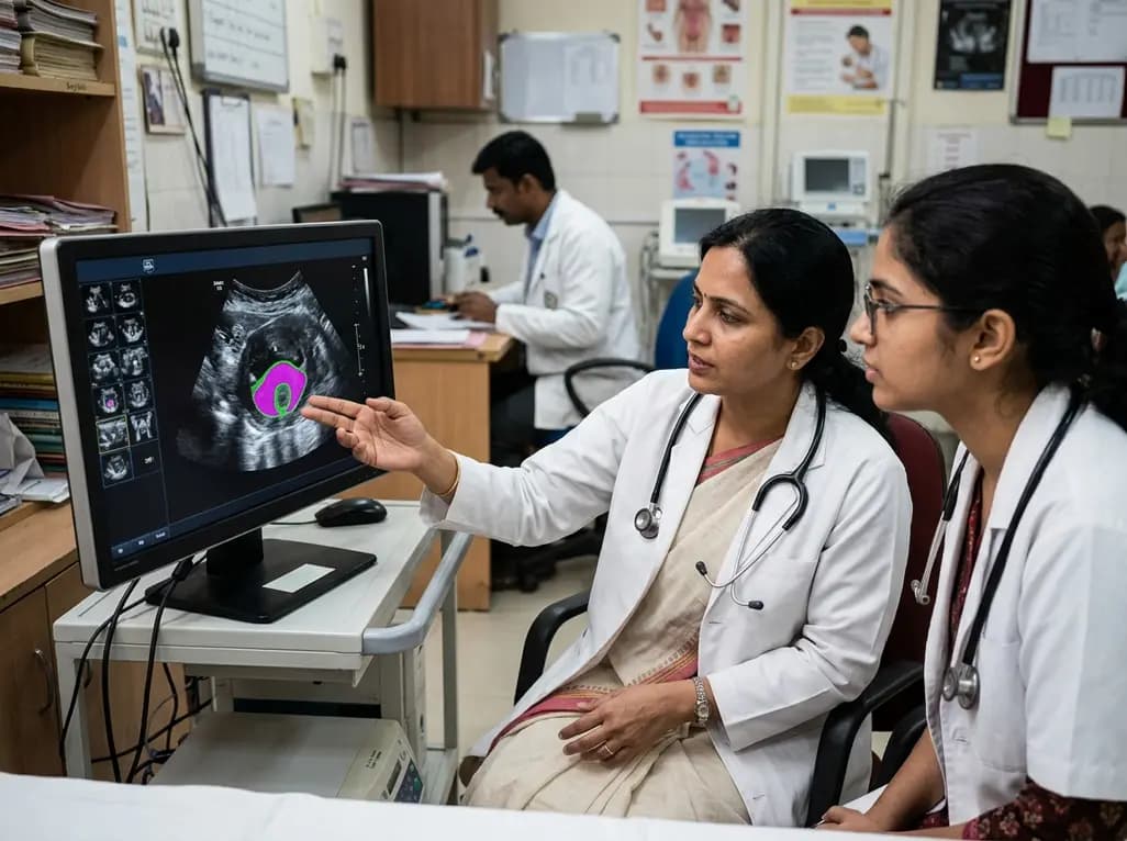
New evidence evaluates MoUNets, a static-weighted ensemble of UNet experts for automated levator hiatus segmentation in pelvic floor ultrasound. Learn how deep learning architectures perform in pelvic organ prolapse diagnostics and clinical practice.
Today
Managing destructive Citrobacter koseri endocarditis with aortocavitary fistula requires meticulous antiplatelet bridging, extensive debridement, and complex redo aortic root and RVOT reconstruction.
Today

A cross-sectional study reveals that elevated METS-IR is independently associated with higher myopia odds in adolescents, showing biological synergy with obesity and highlighting the need for metabolic screening in pediatric eye care.
Today
