.webp&w=384&q=75)

Loading, please wait...
Loading, please wait...

Recent neurodegenerative research highlights how neuroinflammation and microglial activation drive Lewy body disease progression. Pathologists and neurologists long recognized intraneuronal alpha-synuclein accumulation as the defining feature of Lewy body disorders. However, researchers now focus on non-neuronal immune mechanisms in specific hippocampal circuits to explain clinical variability and neurodegeneration.
Histopathologic staging models traditionally emphasize neuronal alpha-synuclein deposition across distinct anatomical regions. However, classic staging frameworks often overlook regions with direct synaptic connectivity, such as the intrahippocampal circuit. Consequently, investigators evaluated post-mortem brain tissue to clarify how microglial processes interact with synaptic pathology. The study examined autopsy-confirmed cases with minimal age-related copathologies to isolate specific neurodegenerative changes. Furthermore, researchers compared these cases against primary age-related tauopathy controls to account for amyloid-independent tau accumulation. By measuring specific marker coverage across six hippocampal subfields, the investigators established clear correlations between neuroinflammation and pathology.
Specifically, the data indicate that microglial activation closely tracks synuclein accumulation in interconnected subfields. Consequently, incorporating regional microglial metrics into existing frameworks significantly enhances our understanding of disease dynamics. Neurologists and pathologists can now evaluate how inflammatory responses parallel synaptic disruption. Ultimately, these biological insights provide a clearer perspective on how structural connectivity influences local neuroinflammation and accelerates clinical dysfunction.
Digital histology techniques provided unprecedented resolution when assessing microglial morphologic and proteomic phenotypes across hippocampal regions. Interestingly, the cornu ammonis 2 subfield demonstrated the highest density of activated microglial markers in patients with synuclein pathology. While total microglial density measured by Iba1 showed baseline presence, activation-specific markers displayed significant elevations.
Furthermore, linear mixed-effects models confirmed that these regional differences remained significant after controlling for demographic variables. Patients exhibited pronounced microglial reactivity in the CA2 subfield compared to other hippocampal regions. Consequently, researchers identified CA2 as an epicenter for neuroinflammatory activity in Lewy body disorders. This subfield appears uniquely vulnerable to pathological stress, serving as a focal hub for local immune activation. In addition, these findings demonstrate that microglial responses are highly regional rather than uniformly distributed throughout the brain. Thus, subfield-specific analysis remains vital when investigating central nervous system pathology and structural neurodegeneration. Neurologists should appreciate how localized immune responses modulate disease trajectories.
Quantitative analysis revealed striking differences in specific microglial proteomic activation markers between patient cohorts. Patients with synuclein accumulation showed marked increases in HLA-DR and CD68 percent area occupied across hippocampal subfields. Specifically, HLA-DR indicates antigen presentation capabilities, whereas CD68 reflects lysosomal activity and phagocytic engagement.
However, general microglial markers like Iba1 did not correlate directly with alpha-synuclein load in the CA2 subfield. Instead, elevated HLA-DR and CD68 levels correlated tightly with local synuclein pathology. Therefore, phenotypic activation states offer far greater diagnostic precision than overall microglial cell counts. In addition, these proteomic shifts reflect active cellular engagement with neurodegenerative pathology rather than passive bystander responses. Pathologists can utilize these distinct marker patterns to differentiate active neuroinflammation from resting microglial states. Consequently, targeting specific microglial activation markers could inform future diagnostic panel design and therapeutic strategies aimed at modulating central neuroinflammation. Understanding these cellular phenotypes enhances clinical assessment of disease severity.
To evaluate pathological spread within the intrahippocampal circuit, researchers divided patients into distinct anatomical subtypes. The Focal Subtype exhibited synuclein pathology restricted to CA2 and CA3 subfields. Conversely, the Widespread Subtype demonstrated broad pathology extending across additional hippocampal regions. Notably, patients in the Widespread Subtype displayed significantly higher CA2 HLA-DR and CD68 levels alongside more severe cognitive impairment.
Furthermore, statistical analysis uncovered a crucial spatial relationship regarding pathological spread. CA2 microglial activation markers correlated strongly with distal alpha-synuclein load in retrogradely connected subfields, but not in anterogradely connected regions. Consequently, these findings suggest that microglial activation closely links to retrograde synaptic transmission of synuclein pathology. Indeed, microglial states may actively facilitate or respond to retrograde axonal transport of toxic synuclein species. Thus, mapping synaptic connectivity alongside neuroinflammatory markers provides invaluable insights into disease propagation mechanics throughout neural circuits. Neurologists can leverage this concept to better predict spatial progression patterns in clinical settings.
Integrating microglial phenotypic markers into histopathological models represents a major advancement in neurodegenerative disease staging. Traditionally, staging relied almost exclusively on neuronal pathology, ignoring active glial contributions to neurodegeneration. However, this study demonstrates that microglial activation states directly track clinical severity and cognitive decline in affected individuals.
In addition, identifying subfield-specific inflammation offers promising opportunities for targeted biomarker development. Clinicians may eventually utilize specialized fluid or imaging biomarkers reflecting microglial activation to monitor disease progression in living patients. Furthermore, understanding the precise timing of microglial activation relative to synuclein spread could highlight new therapeutic windows. Interventions targeting specific microglial phenotypes might slow retrograde transmission and preserve cognitive function. Consequently, combining neuropathological staging with neuroinflammatory profiling will refine diagnostic models and enhance personalized treatment approaches for Lewy body disorders. Ultimately, these advancements bridge basic neuropathology with meaningful clinical outcomes.
Modulating microglial function represents a promising avenue for disease-modifying therapies in Lewy body disorders. Because microglial phenotypes range from protective homeostatic states to destructive pro-inflammatory profiles, therapeutic strategies must be carefully tailored. Specifically, non-selective immunosuppression may fail, whereas targeted inhibition of damaging activation pathways could prove highly effective.
Furthermore, evaluating CA2 microglial activity provides a concrete biochemical readout for pre-clinical drug testing. Researchers can evaluate whether novel drug candidates reduce HLA-DR and CD68 expression while preserving synaptic integrity. In addition, future studies should investigate how co-occurring neuropathologies influence regional microglial reactivity. Understanding these multi-factorial interactions will enable clinicians to optimize patient selection for neuroprotective clinical trials. Consequently, microglial phenotyping stands at the forefront of translational research in neurodegenerative medicine, offering new hope for clinical interventions.
The hippocampal CA2 subfield acts as a focal hub for neuroinflammation in Lewy body disease. Research shows that CA2 exhibits the highest concentration of activated microglial markers, such as HLA-DR and CD68. Furthermore, microglial reactivity in CA2 correlates strongly with alpha-synuclein load and retrograde pathological spread across intrahippocampal circuits. Consequently, CA2 microglial phenotyping provides vital insights into clinical severity and cognitive decline in affected patients.
While Iba1 measures overall microglial presence and cell density, HLA-DR and CD68 specifically reflect functional microglial activation states. HLA-DR indicates antigen presentation capabilities, whereas CD68 marks lysosomal activity associated with active phagocytosis. In Lewy body disease, activated markers like HLA-DR and CD68 correlate directly with local alpha-synuclein accumulation, whereas total Iba1 expression does not. Thus, phenotypic activation markers offer far superior diagnostic precision for tracking active neuroinflammation.
Activated microglial phenotypes in the CA2 subfield correlate specifically with distal alpha-synuclein accumulation in retrogradely connected brain subfields, rather than anterogradely connected regions. This spatial correlation indicates that microglial activation closely tracks or facilitates retrograde synaptic transmission of pathology along neural circuits. Consequently, patients displaying widespread synuclein spread and elevated CA2 microglial activation experience significantly greater cognitive impairment, highlighting the clinical relevance of this immune response.
Disclaimer: This content is for informational and educational purposes only and does not constitute medical advice. Refer to the latest local and national guidelines for clinical practice.
References
1. Luna E et al. Activated microglial phenotypes in the hippocampal CA2 subfield are implicated in Lewy body disease progression. Acta Neuropathol. 2026 Aug 08. doi: undefined. PMID: 42570991.
2. Akiyama H et al. Inflammation and Alzheimer's disease. Neurobiol Aging. 2000;21(3):383-421.

Read summarized clinical updates, watch expert medical content, and earn CME certifications right from your smartphone.
Open your smartphone camera to scan and install.





A new study reveals that activated microglial phenotypes in the hippocampal CA2 subfield, particularly HLA-DR and CD68 markers, are strongly linked to retrograde alpha-synuclein transmission and cognitive decline, offering novel insights into Lewy body disease progression.
last month
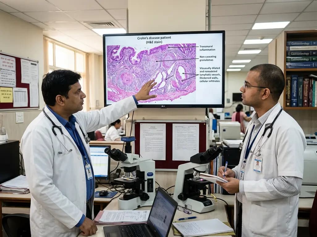
Intestinal lymphatic dysfunction is re-emerging as a fundamental driver of Crohn's disease pathogenesis. Explore how structural remodeling, lymphangitis, and defective clearance shape transmural inflammation and offer novel therapeutic targets.
Today

A school-based intervention using the Health Action Process Approach (HAPA) significantly enhanced adolescents' perceived benefits of consulting healthcare providers and promoted proactive menstrual health behaviors, offering key clinical insights for managing adolescent dysmenorrhea and endometriosis.
Today

A comprehensive analysis of the Translational Maturity Framework for nanomedicine-device combination products, evaluating regulatory complexity, manufacturing hurdles, and clinical readiness across oncology, cardiology, and orthopedics.
Today

A cross-sectional study reveals that sarcopenic obesity independently increases functional impairment and severe disc degeneration in patients with degenerative lumbar spinal stenosis, highlighting the need for body composition assessment.
Today

A landmark dose-response meta-analysis of 8.8 million adults reveals that high ultra-processed food intake significantly elevates risks for cardiovascular disease, cancer, diabetes, depression, and all-cause mortality, highlighting urgent needs for clinical lifestyle interventions.
Today
