

Loading, please wait...
Loading, please wait...

In modern oncology, pathology reports deliver far more than a basic confirmation of invasive malignancy. Specifically, breast cancer biomarker accuracy governs every critical systemic therapy decision. Routine evaluation of hormone receptors, HER2 expression, and Ki-67 indices dictates modern therapeutic pathways. Furthermore, the clinical emergence of the HER2-low category has radically elevated the analytical threshold for diagnostic precision. Slight antigen degradation can mistakenly reclassify an eligible patient, denying them life-prolonging antibody-drug conjugate therapy. Consequently, multidisciplinary teams cannot view biomarker testing as an isolated laboratory procedure. Reliable molecular profiling depends entirely on tissue integrity before technical staining begins. If cells undergo autolysis in the surgical theater, subsequent assays cannot restore lost antigenic sites. Therefore, maintaining cellular quality during the initial surgical phase serves as an indispensable prerequisite for individualized care. Surgical teams must recognize that diagnostic precision begins right at the operating table. In modern oncology, comprehensive tissue management protects patient outcomes across all clinical settings. Additionally, false-negative test results compromise endocrine therapy selection, exposing patients to unnecessary cytotoxic chemotherapy toxicities. Clinicians who understand these biological risks can proactively implement protective measures across institutional boundaries. Preserving vulnerable proteins requires relentless vigilance throughout each phase of surgical transfer and processing.
Clinical audits identify four recurrent, preventable failures that routinely undermine analytical reliability across diverse surgical institutions. First, surgical teams frequently leave excised breast specimens unfixed in operating theaters during prolonged surgical reconstructions. In these warm environments, cellular hypoxia triggers immediate enzymatic self-digestion and rapid protein degradation. Second, specimens from late-Friday or weekend surgeries frequently sit unattended because surgical facilities lack standardized after-hours pathology receiving protocols. Consequently, tissue languishes in transport containers without timely formal accessioning or temperature control. Third, teams often mismanage large lumpectomy and mastectomy specimens through inadequate gross dissection and insufficient fixative volumes. When handlers submerge large, intact surgical specimens into small volumes of formalin, the fixative cannot penetrate dense parenchymal tissue effectively. As a result, the tumor core undergoes extensive autolysis while outer margins fix unevenly. Fourth, surgical units regularly schedule oncologic resections without providing advance notification to the pathology department. Because laboratory staff cannot anticipate incoming specimens, personnel cannot prepare fresh reagents or adjust grossing schedules. Furthermore, this lack of communication generates avoidable delays in specimen bisection. Addressing these four distinct bottlenecks effectively eliminates the primary drivers of tissue degradation in routine hospital practice.
Cold ischemia time refers to the interval between vascular devascularization or surgical tissue excision and immersion in chemical fixative. International oncology guidelines consistently mandate that clinical teams keep cold ischemia time strictly below sixty minutes. When ischemia extends beyond one hour, hydrolytic intracellular enzymes degrade phosphorylated proteins and destabilize delicate hormone receptors. Progesterone receptors prove particularly vulnerable to ischemic delays, showing dramatic signal loss after prolonged bench exposure. Similarly, HER2 protein epitopes undergo conformational alterations that impede antibody binding during immunohistochemistry. Once tissue enters fixative, chemical parameters require equally rigorous oversight. Pathologists universally designate ten percent neutral buffered formalin as the single gold standard fixative for breast oncology. However, formalin penetrates tissue at an average speed of merely one millimeter per hour. Therefore, pathologists must slice large breast specimens at five-millimeter intervals to permit uniform chemical penetration throughout the entire tumor mass. Moreover, total fixation time must remain strictly between six and seventy-two hours. Fixation under six hours leads to inadequate protein cross-linking and tissue distortion. Conversely, over-fixation exceeding seventy-two hours causes irreversible epitope masking. Maintaining these strict chemical boundaries guarantees reproducible testing.
To overcome operational variability, healthcare institutions must implement a standardized, multidisciplinary specimen handling checklist across operating suites and pathology laboratories. This practical checklist formally assigns individual accountability for specimen identification, packaging, transfer, fixation, and digital documentation. Immediately upon excision, the circulating nurse records the exact surgical devascularization time in the electronic operative register. Concurrently, the surgical team verifies patient identifiers, specimen orientation, and lesion characteristics before initiating specimen transfer. Operating staff must transport specimens immediately to the pathology accession desk rather than waiting for formal operative closure. In addition, hospitals need explicit operational pathways for late-afternoon, Friday, and weekend procedures. Dedicated on-call pathology technicians must receive, weigh, ink, and bread-loaf large resections before initiating formalin immersion. Pathologists and laboratory technicians must systematically record the volume ratio of formalin to tissue, targeting a minimum ten-to-one proportion. Furthermore, laboratories must record the exact start and end times of chemical fixation on grossing sheets. Standardizing these logistical handoffs eliminates ambiguity and bridges the traditional gap between surgical theaters and diagnostic suites. Ultimately, structured institutional checklists transform quality assurance from an abstract ambition into verifiable daily clinical practice.
Pre-analytical errors generate devastating clinical and economic consequences, particularly across healthcare networks in developing and resource-limited settings. When suboptimal tissue preservation degrades receptor proteins, laboratory testing generates false-negative results. Consequently, medical oncologists may erroneously withhold highly effective hormonal therapies like tamoxifen or aromatase inhibitors from patients with hormone-responsive disease. In addition, falsely negative HER2 results prevent patients from receiving targeted monoclonal antibodies or cutting-edge antibody-drug conjugates. Conversely, inadequate fixation can produce irregular edge artifacts and false-positive membrane staining, unnecessarily exposing patients to expensive and cardiotoxic therapies. Furthermore, repeat biopsies and reflex molecular testing impose substantial financial burdens on vulnerable healthcare systems and families. In many regional hospitals, surgical units operate far from centralized pathology centers, compounding transport delays and temperature fluctuations. Establishing regional transport protocols, such as refrigerated transfer containers and standardized cold chains, provides practical protection against tissue degradation. Clinicians must realize that sophisticated automated stainers and advanced gene sequencing platforms cannot overcome degraded biological specimens. Therefore, prioritizing disciplined surgical handling represents the most cost-effective strategy to protect precision oncology and elevate breast cancer outcomes globally.
Cold ischemia begins the instant surgeons clamp or transect the tumor vascular supply. Within minutes of devascularization, tissue hypoxia activates intracellular autolytic enzymes that rapidly degrade fragile target proteins and nucleic acids. Progesterone receptors and phosphorylated epitopes prove especially vulnerable to ischemic decay. Restricting cold ischemia to under sixty minutes guarantees that tumor antigens remain intact for immunohistochemistry, preventing false-negative interpretations that could derail life-saving systemic therapies.
Both under-fixation and over-fixation severely distort diagnostic accuracy. Inadequate fixation lasting under six hours leaves cellular proteins under-crosslinked, allowing tissue architecture to collapse and causing false-negative staining during processing. Conversely, over-fixation extending beyond seventy-two hours creates excessive methylene cross-links that permanently mask antigenic epitopes. Both scenarios impair antibody binding during immunohistochemistry, causing inaccurate hormone receptor scoring and misclassification of HER2 expression, which deprives patients of appropriate targeted therapies.
Surgical teams operating outside routine hours must establish designated handover pathways with the pathology department. Operating staff should notify laboratory personnel before initiating surgery. Once the specimen is excised, trained personnel must promptly transport it to the laboratory, where staff slice the specimen at five-millimeter intervals to ensure adequate formalin penetration. If grossing personnel are unavailable, teams must record the cold ischemia time and place bread-loaved tissue into sufficient fixative without delay.
Disclaimer: This content is for informational and educational purposes only and does not constitute medical advice, diagnosis, or treatment. Healthcare professionals should exercise their independent clinical judgment. Refer to the latest local and national guidelines for clinical practice.
References
Paroneto B et al. Before the Pathology Report: Pre-analytical Factors Affecting Biomarker Accuracy in Breast Cancer. Cureus. 2026 Nov undefined. doi: 10.7759/cureus.120033. PMID: 43068044.
Wolff AC, Somerfield MR, Dowsett M, et al. Human Epidermal Growth Factor Receptor 2 Testing in Breast Cancer: ASCO-College of American Pathologists Guideline Update. J Clin Oncol. 2023;41(22):3867-3872.
Allison KH, Hammond MEH, Dowsett M, et al. Estrogen and Progesterone Receptor Testing in Breast Cancer: ASCO/CAP Guideline Update. J Clin Oncol. 2020;38(12):1346-1366.

Read summarized clinical updates, watch expert medical content, and earn CME certifications right from your smartphone.
Open your smartphone camera to scan and install.





Pre-analytical handling directly dictates the reliability of ER, PR, HER2, and Ki-67 testing in breast cancer. This review outlines how controlled ischemia, standardized formalin fixation, and structured operating room communication prevent diagnostic failures and protect precision oncology decisions.
Today
A groundbreaking Bayesian two-stage clinical trial design integrates historical reference data using MAP priors to evaluate biosimilarity and interchangeability efficiently, minimizing patient burden in rheumatoid arthritis studies.
Today
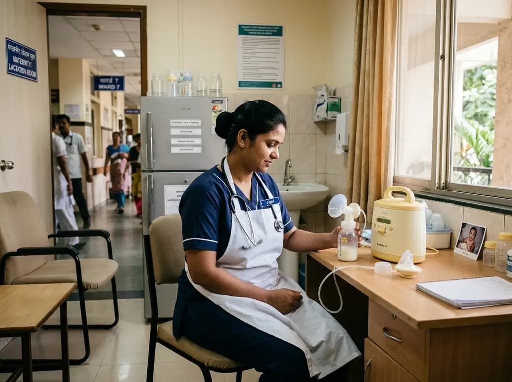
A qualitative study in the Journal of Human Lactation highlights the significant hurdles healthcare workers face when continuing breastfeeding. Inadequate facilities, rigid shift schedules, and lack of institutional policies demand structured workplace lactation support and compliant hospital infrastructure.
Today
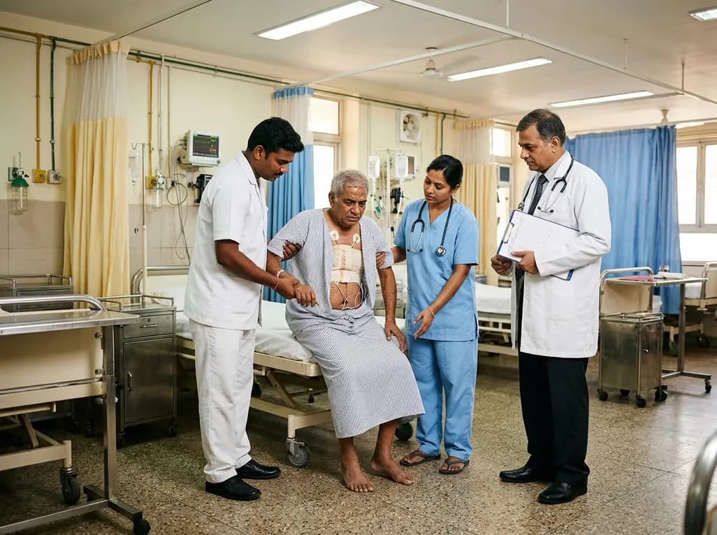
A cross-sectional study evaluates the critical intersection between acute surgical pain, kinesiophobia, and fall risks during initial mobilization after cardiac surgery, emphasizing targeted nursing strategies.
Today

A recent cross-sectional analysis reveals that over a quarter of randomized controlled trials for non-specific low back pain face discontinuation or unknown status, while more than 36% of completed trials remain unpublished, exposing notable reporting gaps in spine and musculoskeletal clinical research.
Today
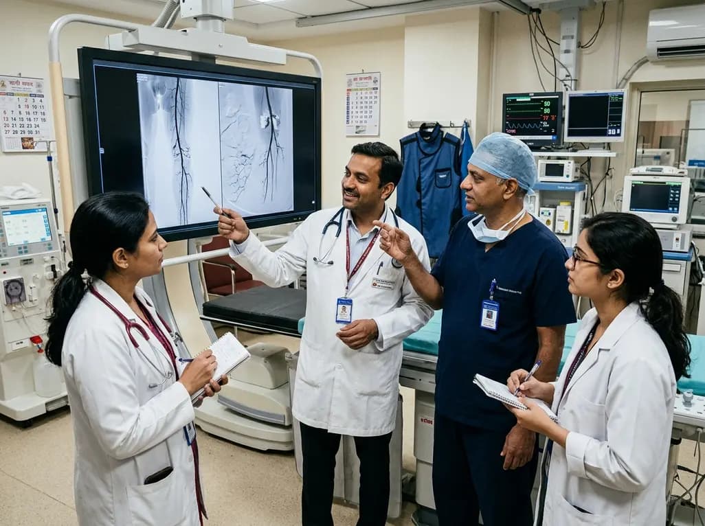
Explore revascularisation strategies in dialysis-dependent chronic limb-threatening ischaemia. This review examines surgical bypass vs endovascular therapy, BEST-CLI trial findings, procedural morbidity, conduit availability, and multidisciplinary decision-making for optimal limb salvage.
Today
