

Loading, please wait...
Loading, please wait...

Central nervous system inflammatory disorders present distinct diagnostic challenges, therapeutic pathways, and neurodegenerative profiles. Evaluating long-term brain atrophy in MOGAD is essential because clinicians frequently struggle to differentiate this antibody-mediated entity from classic multiple sclerosis. While multiple sclerosis causes insidious and continuous global tissue loss, clinicians now recognize that antibody-associated syndromes follow different injury dynamics. Consequently, understanding regional structural changes helps neurologists tailor immunotherapy, preserve parenchymal volume, and predict disability outcomes effectively.
Multiple sclerosis (MS), myelin oligodendrocyte glycoprotein antibody-associated disease (MOGAD), and aquaporin-4 antibody-positive neuromyelitis optica spectrum disorder (AQP4+ NMOSD) involve distinct cellular targets. Specifically, MS triggers widespread perivenular demyelination along with chronic compartmentalized inflammation in the meninges and normal-appearing white matter. This ongoing cascade causes diffuse neuroaxonal degradation over time. Conversely, AQP4+ NMOSD represents an autoimmune astrocytopathy where antibody binding causes complement-mediated astrocyte destruction, secondary oligodendrocyte injury, and necrosis.
In sharp contrast to both conditions, MOGAD primarily damages the outer lamellae of myelin sheaths through transient autoantibody responses. Oligodendrocytes frequently survive these acute inflammatory insults. As a result, tissue recovery often occurs after relapses, leading to lesion resolution on neuroimaging. However, clinicians historically questioned whether MOGAD patients experience silent neurodegeneration similar to MS progression. Understanding these divergence points requires rigorous longitudinal morphometric investigations. Therefore, researchers sought to isolate whether brain volume reduction in antibody-positive disorders occurs continuously or solely following overt acute attacks.
To characterize these structural trajectories, investigators within the Magnetic Resonance Imaging in Multiple Sclerosis (MAGNIMS) network executed a robust multicenter study across fifteen specialized centers. The study included adult patients diagnosed with MS, MOGAD, or AQP4+ NMOSD alongside healthy controls. Importantly, each eligible participant completed at least two brain magnetic resonance imaging scans on the exact same scanner. Furthermore, the protocol mandated that serial scans occurred at least six months apart and at least three months post-relapse. This design specifically eliminated acute inflammatory edema and pseudoatrophy confounds.
Researchers segmented white matter lesions using the automated Lesion Segmentation Toolbox. Additionally, they performed volumetric quantification on lesion-filled T1-weighted images via Sequence Adaptive Multimodal Segmentation using within-subject template approaches. The team classified MOGAD lesion dynamics into stable lesions, new lesion formation, or lesion resolution. Subsequently, investigators applied linear mixed-effects models adjusted for age, biological sex, baseline parenchymal volume, and MRI center. Consequently, this stringent methodological framework provided exceptional precision when evaluating subtle regional and global parenchymal shifts across distinct neuroinflammatory cohorts.
The study examined 489 individuals, comprising 98 patients with MS, 72 with MOGAD, 41 with AQP4+ NMOSD, and 278 healthy controls. The mean follow-up period spanned 2.44 years for MS, 2.57 years for MOGAD, and 3.22 years for NMOSD. Compared to healthy controls, MS demonstrated accelerated annualized global brain volume loss. In contrast, patients with MOGAD did not display generalized cortical thinning. Instead, they exhibited selective white matter volume reduction and progressive ventricular expansion, notably within the lateral and third ventricles.
Meanwhile, individuals with AQP4+ NMOSD showed distinct pallidal volume reduction without accelerated global cortical shrinkage. Furthermore, phenotypic stratification within the MOGAD cohort uncovered profound pathophysiological insights. Approximately 62.5% of MOGAD participants experienced a relapsing disease course. Remarkably, these relapsing individuals exhibited significantly greater third ventricular enlargement than patients with MS. Moreover, MOGAD patients with newly accrued lesions displayed significantly faster lateral and third ventricular expansion compared to healthy controls, MS, and NMOSD. Conversely, patients with stable scans or resolving lesions showed preserved ventricular dimensions. Thus, brain atrophy in MOGAD reflects episodic inflammatory activity rather than relentless neurodegeneration.
These morphometric observations establish a critical distinction in central nervous system pathology. In multiple sclerosis, brain atrophy develops continuously throughout the disease course. Microglial activation, chronic oxidative stress, and smoldering compartmentalized inflammation drive insidious neuroaxonal destruction even during clinical remission. Therefore, progressive whole-brain and gray matter volume loss occurs independently of relapse activity in MS.
Conversely, tissue damage in MOGAD follows an event-driven injury model. Because oligodendrocytes survive many inflammatory episodes, substantial remyelination and lesion resolution remain frequent. However, when acute relapses or newly accrued focal white matter lesions emerge, severe focal perivascular damage occurs. This destructive focal insult severs traversing axonal tracts. Consequently, secondary Wallerian degeneration causes periventricular white matter thinning and subsequent ventricular dilation. Because ventricular expansion tracks new lesion accrual rather than clinical duration, tissue loss in MOGAD directly reflects focal inflammatory episodes. In addition, this mechanistic insight proves that MOGAD lacks the silent, relentless neurodegenerative component seen in classical MS.
These imaging findings carry direct clinical ramifications for practicing neurologists and radiologists evaluating demyelinating disease. In daily clinical management, treating physicians should not automatically apply MS monitoring paradigms to patients diagnosed with MOGAD. For instance, while accelerated global brain atrophy serves as a validated surrogate endpoint for neurodegeneration in MS clinical trials, it lacks equal clinical utility in MOGAD. Instead, monitoring new lesion development and progressive central ventricular expansion provides superior clinical insight into subclinical inflammatory activity.
Furthermore, these findings emphasize the paramount necessity of preventing clinical and radiological relapses in MOGAD. Because neuroaxonal injury accumulates during active inflammatory breakthroughs, timely escalation of maintenance immunotherapy remains crucial. Maintenance agents such as intravenous immunoglobulin, rituximab, or azathioprine can prevent permanent structural injury when deployed effectively. Consequently, clinicians should monitor serial imaging for subtle ventricular dilation and lesion recurrence. By halting acute inflammatory breakthroughs, neurologists directly arrest the primary driver of irreversible brain tissue loss in these patients.
Although this multicenter study delivers vital insights, clinicians must contextualize several methodological limitations. First, the AQP4+ NMOSD cohort remained relatively modest in size, reflecting the rarity of this severe astrocytopathy. Second, follow-up durations varied across centers, although linear mixed-effects modeling effectively adjusted for repeated measures and inter-scanner variations. Third, standard T1-weighted volumetry may overlook microscopic damage in normal-appearing brain parenchyma.
Therefore, future research must incorporate advanced imaging techniques to refine diagnostic precision. Specifically, ultra-high-field MRI, diffusion tensor imaging, and serum neurofilament light chain quantification could clarify acute versus chronic axonal compromise. Additionally, prospective studies should evaluate how novel targeted therapeutics influence long-term regional tissue preservation. Ultimately, establishing standardized volumetric neuroimaging protocols will assist multidisciplinary teams in distinguishing MOGAD from MS early, preventing misdiagnosis and optimizing individualized patient management protocols worldwide.
Multiple sclerosis causes diffuse, relentless global gray and white matter atrophy driven by continuous neurodegeneration. In contrast, brain atrophy in MOGAD follows an event-driven trajectory. Tissue loss in MOGAD is primarily restricted to white matter and ventricular enlargement, occurring directly downstream of acute focal inflammatory relapses and new lesion formation.
New focal lesions in MOGAD trigger focal tissue destruction and subsequent secondary Wallerian tract degeneration. As axonal fibers traveling through periventricular white matter tracts deteriorate, structural volume decreases. Consequently, the brain ventricles expand, making lateral and third ventricular enlargement reliable structural surrogates of cumulative focal inflammatory activity.
This differentiation dictates therapeutic strategy and prognosis. Progressive neurodegeneration in multiple sclerosis requires neuroprotective and disease-modifying agents targeting chronic compartmentalized inflammation. Conversely, preventing brain tissue loss in MOGAD relies entirely on suppressing acute relapses and new focal lesion accrual using targeted maintenance immunotherapies, such as immunosuppressants or intravenous immunoglobulins.
Disclaimer: This content is for informational and educational purposes only. It is not intended to substitute for professional medical advice, diagnosis, or treatment. Always seek the advice of your physician or other qualified healthcare provider with any questions you may have regarding a medical condition. Refer to the latest local and national guidelines for clinical practice.
References

Read summarized clinical updates, watch expert medical content, and earn CME certifications right from your smartphone.
Open your smartphone camera to scan and install.





A multicenter MAGNIMS study reveals that brain atrophy in MOGAD follows an event-driven trajectory driven by acute relapses and new lesions, unlike the continuous diffuse neurodegeneration in multiple sclerosis. These insights highlight the vital role of relapse prevention.
Today

Review how tailored exercise programs benefit frail older patients with heart failure. Explore pathophysiological mechanisms, individualized training prescriptions, and strategies to overcome adherence barriers in clinical practice.
Today
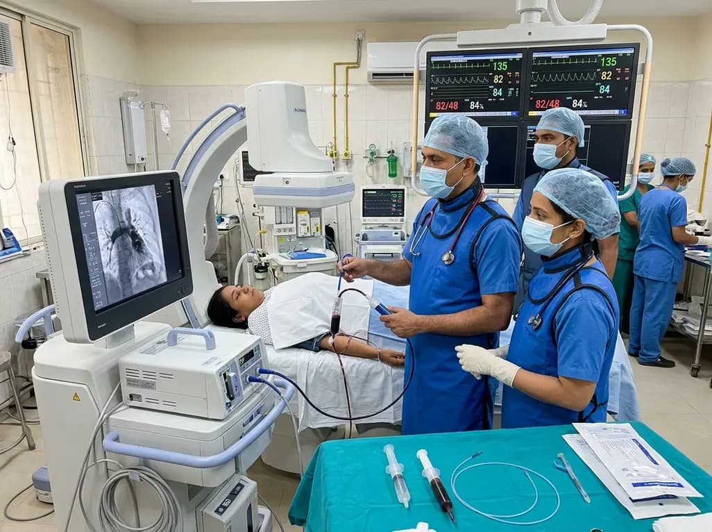
In high-risk pulmonary embolism complicated by severe anemia and active uterine bleeding, systemic thrombolysis is contraindicated. Large-bore aspiration thrombectomy provides rapid hemodynamic rescue without fibrinolysis, supported by inferior vena cava filter placement and multidisciplinary critical care.
Today

A cross-sectional study in Zanzibar reveals that 46.5% of adults with type 2 diabetes experience multidomain autonomic dysfunction on the COMPASS-31 tool, primarily orthostatic intolerance, while 45.9% exhibit cardiovascular autonomic neuropathy, highlighting the urgent need for structured clinical screening.
Today

This comparative study evaluates posterior labral tears with and without glenohumeral instability, highlighting distinct demographic patterns, MRI underestimation in stable shoulders, and successful arthroscopic repair outcomes.
Today

A recent case-control study reveals that urinary clusterin provides superior diagnostic performance for early diabetic kidney disease detection in pediatric type 1 diabetes mellitus, identifying tubular damage before conventional microalbuminuria occurs.
Today
