

Loading, please wait...
Loading, please wait...
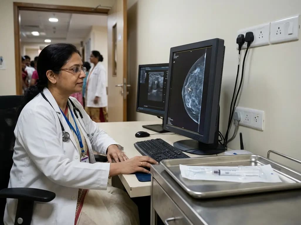
Breast tissue markers have emerged as an indispensable cornerstone of modern breast cancer care and interventional oncology. Recently, global medical technology leader BD officially introduced its advanced UltraCor Twirl breast tissue markers in India. This launch addresses a critical clinical need across diagnostic radiology and surgical oncology practices. Specifically, breast interventions require dependable anatomical landmarks that remain stable across complex diagnostic pathways. Modern treatment protocols increasingly incorporate neoadjuvant systemic therapies, which frequently cause target lesions to diminish prior to definitive surgery. Consequently, reliable localization devices ensure seamless coordination between interventional radiologists, medical oncologists, and surgical teams.
Percutaneous core needle and vacuum-assisted breast biopsies represent standard diagnostic procedures for suspicious lesions. However, removing targeted tissue frequently alters the local microarchitecture or evacuates small calcification clusters entirely. Under these circumstances, breast tissue markers provide an indelible visual reference for subsequent clinical evaluation. Historically, early metallic clips suffered from technical limitations, including post-biopsy displacement and poor visibility on ultrasound. Therefore, medical device manufacturers invested heavily in advanced metallurgic engineering to resolve these procedural vulnerabilities.
Additionally, the rapid expansion of breast-conserving surgery demands heightened spatial precision from the initial diagnostic encounter. When pathology reveals invasive carcinoma or ductal carcinoma in situ, clinicians must accurately correlate initial imaging abnormalities with surgical margins. Breast tissue markers establish this vital link. Furthermore, implanting a permanent fiducial marker enables serial radiographic tracking during multi-cycle systemic therapies. By establishing a definitive reference point at the biopsy site, multidisciplinary care teams eliminate diagnostic ambiguity and optimize surgical margins. Moreover, modern guidelines consistently advocate immediate marker placement during any image-guided biopsy where tissue remodeling could obscure subsequent localization.
The UltraCor Twirl introduces specialized mechanical characteristics engineered to overcome the physical limitations of conventional biopsy clips. Primarily, the device incorporates a precision-engineered nitinol wireform that forms a three-dimensional ring geometry upon deployment. Because nitinol possesses unique superelasticity and thermal shape memory, the implant smoothly traverses the rigid 17-gauge delivery needle. It then immediately springs into its predefined circular shape within the biopsy cavity.
Furthermore, the device utilizes an innovative polymer-free and gel-free architecture. Traditional markers often embed metallic components within collagen, cellulose, or bioabsorbable synthetic polymers to facilitate temporary sonographic visibility. However, these organic carriers gradually degrade, occasionally leading to delayed clip movement or transient inflammatory responses. In contrast, the self-incorporating metallic framework of the UltraCor Twirl engages directly with adjacent breast parenchyma. As a result, the surrounding fibrous stroma integrates rapidly around the nitinol coils. This permanent mechanical fixation maintains long-term positional stability without invoking foreign body granulomatous reactions. Additionally, the pre-loaded delivery applicator features distinct centimeter depth indicators and a positive-locking plunger system. Consequently, interventionists achieve predictable deployment tactile feedback, preventing premature extrusion or misplaced marker deposition during demanding biopsies.
Biopsy clip migration represents a well-documented technical failure that can misdirect downstream surgical interventions and compromise clinical outcomes. Migration typically occurs immediately following needle withdrawal due to negative pressure decompression or local hematoma expansion. However, the three-dimensional geometry of the UltraCor Twirl provides superior anchoring resistance against fluid currents and shear stresses. Consequently, clinical evaluations demonstrate exceptional post-deployment stability without measurable spatial displacement over prolonged observational intervals.
Equally important is the device's uncompromised visibility across multiple complementary imaging modalities. Digital mammography and tomosynthesis easily detect the radiopaque nitinol alloy against dense fibroglandular parenchyma. Furthermore, the specialized coiled shape produces pronounced acoustic interfaces that stand out crisply during high-resolution ultrasound examination. This capability offers a substantial procedural advantage, as ultrasound guidance facilitates comfortable, radiation-free preoperative wire or seed placement. Similarly, the non-ferromagnetic composition ensures compatibility with magnetic resonance imaging. The marker generates minimal susceptibility artifact, preserving high-resolution visualization of surrounding soft tissues. Thus, radiologists can confidently assess residual tumor margins without encountering disruptive signal voids during post-contrast sequences. Moreover, this robust multimodal visibility provides exceptional convenience when patients transfer between diagnostic facilities utilizing different imaging technologies.
The widespread adoption of neoadjuvant systemic therapy has revolutionized modern breast cancer management, particularly for aggressive molecular subtypes. Modern targeted regimens achieve remarkable rates of pathological complete response, often causing extensive primary tumors and involved nodes to resolve radiologically. Consequently, without a reliable fiducial marker placed prior to chemotherapy initiation, surgeons face immense difficulty identifying the original tumor bed.
The UltraCor Twirl marker plays an indispensable role throughout this extended therapeutic continuum. By providing durable anchoring, the marker demarcates the exact epicenter of the initial malignancy throughout multiple months of therapy. Furthermore, the technology proves equally valuable in targeted axillary dissection. Placing the marker inside biopsy-proven metastatic axillary lymph nodes before systemic therapy enables selective surgical harvesting after treatment completion. This targeted approach prevents extensive full axillary lymph node dissections, substantially reducing postoperative lymphedema risks. Therefore, dependable marker technology preserves surgical precision, supports oncological safety, and dramatically elevates the long-term functional quality of life.
Successful marker placement requires meticulous procedural execution and comprehensive knowledge of potential clinical risks. Typically, the interventional radiologist advances the 17-gauge applicator coaxially through the existing biopsy cannula or independently under direct ultrasound guidance. Once the beveled tip reaches the target cavity, the clinician disengages the locking plunger to release the nitinol ring. Following deployment, post-procedure orthogonal mammographic views confirm exact spatial positioning and establish baseline documentation.
Nevertheless, clinicians must observe specific contraindications and safety guidelines during patient selection. Because the UltraCor Twirl consists of a nickel-titanium alloy, clinicians must not implant the device in patients with known nickel hypersensitivity. Additionally, interventionists must exercise caution when operating adjacent to breast implants to avoid accidental puncture of the prosthetic capsule. Proper handling remains equally critical; practitioners should never bend the delivery needle or force the deployment plunger against unexpected anatomical resistance. Furthermore, adhering to strict aseptic technique during every stage of the intervention prevents secondary microbial colonization of the foreign body. Through rigorous adherence to these established clinical safeguards, medical teams consistently ensure maximal diagnostic utility and outstanding patient safety.
Q1: Why is marker placement necessary during a percutaneous breast biopsy?
Percutaneous core biopsies often remove or obscure small suspicious lesions and microcalcifications. Consequently, placing a radiopaque marker preserves the exact anatomical epicenter of the biopsied abnormality. This permanent reference enables multidisciplinary teams to track therapeutic responses accurately. Furthermore, the marker guides future wire or wireless surgical localization if pathology confirms malignancy. Without accurate marking, clinicians face significant challenges in locating the original tumor bed after therapeutic regression.
Q2: How does the nitinol design of UltraCor Twirl prevent marker migration?
The UltraCor Twirl utilizes an elastic nickel-titanium alloy formed into a three-dimensional ring geometry. When clinicians deploy the marker through the 17-gauge needle, the shape memory alloy immediately expands into adjacent breast parenchyma. Therefore, the device anchors firmly into the surrounding tissue bed without requiring chemical polymers or resorbable gels. This mechanical fixation prevents displacement caused by cavity decompression, hematoma formation, or normal glandular tissue movement.
Q3: Can clinicians safely visualize UltraCor Twirl across all imaging modalities?
Yes, clinicians can reliably visualize the UltraCor Twirl marker across digital mammography, tomosynthesis, ultrasound, and magnetic resonance imaging. Notably, the marker's specialized geometry produces distinct echogenicity under ultrasound, eliminating the acoustic shadowing that often obscures standard surgical clips. In addition, the non-ferromagnetic nitinol composition minimizes susceptibility artifacts during breast MRI. Consequently, radiologists can accurately assess tumor regression and plan subsequent breast-conserving interventions throughout every stage of diagnostic and therapeutic follow-up.
Disclaimer: This content is for informational and educational purposes only. It does not constitute medical advice or replace professional judgment. Refer to the latest local and national guidelines for clinical practice.
References

Read summarized clinical updates, watch expert medical content, and earn CME certifications right from your smartphone.
Open your smartphone camera to scan and install.





Medical technology leader BD has introduced the UltraCor Twirl breast tissue marker in India. Engineered from nitinol for multimodal visibility across ultrasound, mammography, and MRI, the device prevents migration and optimizes lesion localization during neoadjuvant chemotherapy and breast-conserving surgery.
Today

With India still adhering to 2009 air pollution benchmarks, the delayed NAAQS revision exposes populations to fine particulate matter far exceeding global safety limits. Clinicians face surging cardiopulmonary morbidity, emphasizing the urgent need for stringent, health-centered regulatory thresholds nationwide.
Today

India's leading assisted reproduction provider Indira IVF is in advanced talks to acquire a majority stake in Kerala's Sabine Hospital in a Rs 1,300 crore transaction. This strategic development highlights rising private equity interest, clinical consolidation, and changing demographic demands across reproductive medicine.
Today

Recent findings from the VESUVE study demonstrate that brain MRI detects intracranial lesions in over 76% of patients with infective endocarditis. Acute ischemic stroke and subarachnoid hemorrhage independently elevate six-month mortality, underscoring the critical prognostic utility of neuroimaging.
Yesterday

Modern 3D anthropometry reveals that pediatric leg growth ceases years before spinal elongation, exposing the clinical illusion of the classical Anderson-Green model. Learn how segmental growth dissociation improves surgical timing for epiphysiodesis in adolescent leg length discrepancy.
Yesterday

A meta-analysis of 18 studies reveals that bariatric surgery does not significantly alter circulating homocysteine levels, regardless of follow-up duration. Clinicians must maintain vigilance over post-surgical micronutrient supplementation to prevent hyperhomocysteinemia.
Yesterday
