.webp&w=384&q=75)

Loading, please wait...
Loading, please wait...
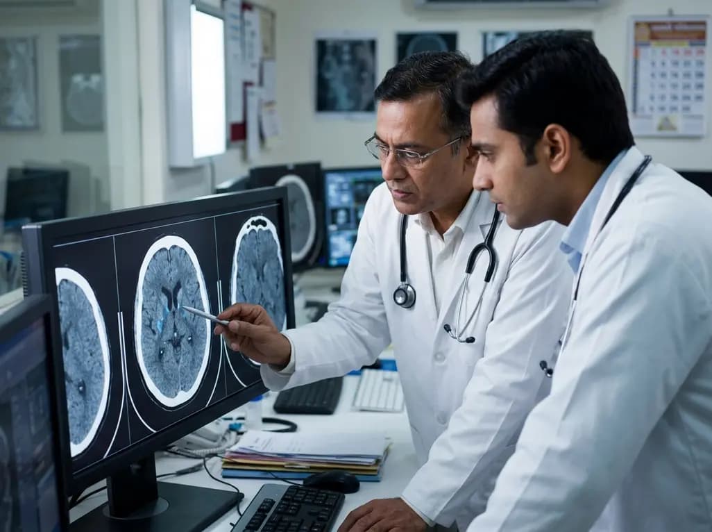
Ischemic events in oncology patients present complex clinical challenges that require prompt identification and multidisciplinary care. Malignancy significantly increases thromboembolic risk through systemic hypercoagulability, direct vascular compression, and endothelial disruption. Consequently, clinicians frequently encounter cancer-associated stroke in patients presenting with acute neurological deficits without traditional vascular risk factors. Identifying robust neuroimaging biomarkers helps clinicians distinguish occult malignancy-related ischemia from conventional arterial disease. Among emerging imaging markers, perivascular spaces—also known as Virchow-Robin spaces—have gained substantial interest in vascular neurology. These fluid-filled structures surround cerebral blood vessels as they course into the brain parenchyma, serving as vital pathways for interstitial fluid drainage and metabolic waste clearance. When perivascular spaces become pathologically enlarged, high-resolution magnetic resonance imaging (MRI) can readily detect them as small punctate or tubular signal abnormalities. While enlarged perivascular spaces in the basal ganglia generally reflect cerebral small vessel disease, their specific relationship with occult malignancy has remained poorly understood. Consequently, clinical researchers have begun investigating whether basal ganglia perivascular alterations might serve as non-invasive indicators of underlying malignancy in acute stroke populations.
To explore this critical diagnostic relationship, researchers conducted a rigorously matched case-control study in Xiamen, China. The investigation recruited 184 acute ischemic stroke patients, dividing them equally into a cancer cohort and a matched non-cancer control group. Patients in both groups underwent baseline high-resolution cranial magnetic resonance imaging upon hospital admission to evaluate neuroimaging abnormalities. Specifically, neuroradiologists systematically graded the severity of enlarged perivascular spaces in the basal ganglia, utilizing validated visual rating scales to ensure objective measurement. In addition to perivascular space assessment, the research team comprehensively collected demographic parameters, clinical risk factors, vascular imaging changes, and admission laboratory findings. Patients' sex distribution showed a male majority of 65.22%, with an overall mean age of approximately 68.83 years across the study population. Subgroup analysis further stratified subjects based on oncology treatment modalities, including surgical intervention, systemic chemotherapy, and radiation therapy. By carefully controlling for traditional stroke risk factors like hypertension, diabetes, and dyslipidemia, the investigators isolated the specific diagnostic contribution of perivascular enlargement in basal ganglia structures.
The study revealed a strong, statistically significant association between basal ganglia perivascular enlargement and stroke in cancer patients. Multivariable logistic regression analysis demonstrated that severe perivascular enlargement independently increased the odds of cancer-associated ischemia by 85%. Specifically, after adjusting for age, gender, cardiovascular risk factors, concomitant neuroimaging markers, and comprehensive blood parameters, the adjusted odds ratio remained highly elevated at 1.85. Furthermore, subgroup analyses demonstrated an even stronger correlation among individuals actively undergoing oncological therapies. Patients receiving active chemotherapy, radiotherapy, or surgical treatments exhibited heightened degrees of basal ganglia perivascular enlargement compared to non-cancer controls. Consequently, these statistical findings suggest that both tumor biological activity and antineoplastic treatments contribute to microvascular glymphatic compromise. Researchers noted that the association was particularly prominent in basal ganglia regions rather than white matter areas like the centrum semiovale. Therefore, localized basal ganglia changes may reflect specific vulnerability to systemic hypercoagulability, microthrombosis, and tumor-induced inflammatory cascades.
To determine whether perivascular imaging could improve clinical detection, researchers constructed multivariate diagnostic prediction models. The primary predictive model integrated basal ganglia perivascular grading alongside admission blood biomarkers, clinical risk profiles, and routine vascular imaging findings. Remarkably, this combined diagnostic model achieved an area under the receiver operating characteristic curve of 0.848. This predictive accuracy proved significantly superior to individual clinical models or isolated imaging parameters alone. Consequently, incorporating basal ganglia perivascular assessment into standard neuroimaging evaluations markedly enhances clinicians' ability to identify underlying hidden malignancies. Furthermore, the high diagnostic yield underscores the clinical utility of routine high-resolution brain MRI scans during initial acute stroke evaluations. When clinicians detect marked basal ganglia perivascular enlargement in stroke patients lacking conventional cardiovascular risk factors, screening for occult cancer becomes highly actionable. Therefore, combining neuroimaging biomarkers with tumor marker panels and systemic staging workups provides a comprehensive strategy for early cancer detection in stroke neurology.
Understanding how cancer leads to perivascular enlargement requires examining the complex interactions between systemic hypercoagulability and glymphatic clearance. Malignancy frequently triggers systemic inflammation, microthrombosis, and elevated blood viscosity through the secretion of procoagulant mucins and tissue factors. Consequently, microvascular stasis and endothelial dysfunction develop within deep subcortical structures, particularly the basal ganglia. Furthermore, impaired glymphatic fluid transit prevents normal clearance of metabolic waste, resulting in fluid accumulation around deep penetrating lenticulostriate arteries. As perivascular spaces progressively enlarge, localized brain tissue perfusion decreases, predisposing vulnerable subcortical regions to ischemic damage. Additionally, antineoplastic medications and radiation therapy can directly induce microvascular injury and blood-brain barrier breakdown. Therefore, perivascular enlargement represents a visible neuroimaging marker of profound systemic vascular toxicity and glymphatic failure. Recognizing this imaging feature allows neurologists and oncologists to initiate prompt diagnostic workups, tailored antithrombotic therapies, and coordinated multidisciplinary management.
The findings from this study open promising avenues for integrating advanced neuroimaging into routine clinical risk stratification. Clinicians managing acute stroke patients should pay close attention to basal ganglia perivascular features, especially when clear vascular risk factors are absent. Furthermore, future prospective clinical trials should evaluate whether targeted occult cancer screening protocols in high-risk stroke patients improve long-term survival and reduce recurrent ischemic events. Standardizing automated magnetic resonance imaging software could also streamline perivascular space quantification across busy clinical centers. Additionally, combining neuroimaging markers with novel liquid biopsy techniques may refine diagnostic precision even further. Ultimately, early identification of cancer-associated ischemia enables timely initiation of appropriate secondary prevention, including low-molecular-weight heparin or direct oral anticoagulants when indicated. As neuro-oncology continues to evolve, incorporating imaging biomarkers like perivascular spaces will play a vital role in optimizing personalized patient care.
Enlarged perivascular spaces are fluid-filled cavities surrounding blood vessels in deep brain structures like the basal ganglia. They become visible on brain magnetic resonance imaging when fluid accumulation occurs, often indicating underlying cerebral small vessel disease, impaired glymphatic drainage, or endothelial injury associated with vascular and systemic conditions.
Cancer increases stroke risk primarily by inducing systemic hypercoagulability through procoagulant tumor factors, mucin secretion, and chronic inflammatory responses. Furthermore, direct tumor compression, endocarditis, and microvascular toxicity from cancer treatments like chemotherapy or radiation therapy further elevate the risk of arterial occlusion and cerebral ischemia.
Evaluating basal ganglia perivascular spaces provides crucial insights into microvascular health and glymphatic function during stroke evaluations. When identified alongside standard clinical risk factors, severe perivascular enlargement helps clinicians suspect occult underlying cancer, guiding timely systemic screening, accurate etiologic classification, and tailored antithrombotic management strategies.
Disclaimer: This content is for informational and educational purposes only and does not constitute medical advice. Healthcare professionals should rely on clinical judgment and refer to official local and national guidelines for clinical practice.
References
Wu J et al. Correlation of enlarged perivascular spaces in basal ganglion and cancer-associated stroke: a case-control study in China. Stroke Vasc Neurol. 2025 Aug 26. doi: 10.1136/svn-2024-003287. PMID: 39532461.
Bang OY et al. Cancer-associated stroke: The meeting point of oncology and neurology. J Stroke. 2020;22(2):189-208.
Wardlaw JM et al. Neuroimaging standards for research into small vessel disease and its contribution to ageing and neurodegeneration. Lancet Neurol. 2013;12(8):822-838.

Read summarized clinical updates, watch expert medical content, and earn CME certifications right from your smartphone.
Open your smartphone camera to scan and install.





A matched case-control study demonstrates that enlarged perivascular spaces in the basal ganglia significantly correlate with cancer-associated stroke (OR=1.85), providing strong diagnostic utility for identifying underlying malignancy in ischemic stroke patients.
2 months ago

Following regulatory scrutiny from FSSAI regarding promotional representations and lean muscle preservation claims, Dr. Reddy's Laboratories and Nestlé Health Science are currently reviewing the branding and marketing strategy for their joint nutritional supplement Celevida GLP+.
Today

Demanding exhaustive hormonal profiling can inadvertently discourage clinical studies involving females. Methodological standards must align with the specific scientific question to expand, not restrict, equitable evidence for women.
Today

The Directorate General of Health Services directs Indian states to halt the hazardous reuse and reprocessing of single-use medical devices. Discover regulatory implications under the Drugs and Cosmetics Act, critical infection risks, legal liabilities, and operational changes required across clinical specialties.
Today

A novel calcium polyphosphate coacervate enables injectable bone implants with in situ hierarchical porosity via phase separation and gas foaming, significantly enhancing cell proliferation, energy metabolism, and osteogenic differentiation for minimally invasive bone defect repair.
Today

India is advancing preventive healthcare by integrating genomics, artificial intelligence, and quantum technologies. With the SANGAM programme sequencing one million Indian genomes, clinicians can unlock personalized therapies, detect cardiometabolic risks early, and curb the burden of diabetes and heart disease.
Today
