

Loading, please wait...
Loading, please wait...

Surgical management of intra-axial tumors within the precentral gyrus presents formidable challenges for modern neurosurgeons. Historically, conventional transcortical corridors required significant gyral incisions that often produced devastating hemiparesis. Consequently, the trans-sulcal approach offers a sophisticated alternative that harnesses natural anatomical corridors to access subcortical pathology. By entering through the sulcal depth, surgeons minimize direct cortical disruption and spare critical motor pathways. In addition, neurosurgeons combine this trajectory with awake functional monitoring to protect motor control. Recent clinical evaluations demonstrate that this strategy achieves optimal cytoreduction while protecting functional mobility.
The precentral gyrus orchestrates voluntary motor execution through densely arranged pyramidal neurons and descending corticospinal tracts. Therefore, even minimal operative traction, direct incision, or thermal injury in this eloquent zone can precipitate irreversible hemiplegia. Infiltrative high-grade gliomas further complicate surgical execution because they obscure normal sulcal boundaries and efface regional anatomy. Furthermore, these invasive tumors displace white matter tracts unpredictably across adjacent motor corridors. Traditional cortical incisions violate functional grey matter directly overlying the mass. Consequently, patients often endure permanent post-resection functional deficits and loss of motor independence. Preserving vascular architecture represents another crucial surgical objective. Specifically, the primary motor cortex depends heavily on delicate pial arterial branches and anastomotic venous networks. Inadvertent injury to these vessels induces local ischemia and secondary tissue necrosis. Thus, surgical access must balance radical cytoreduction against the preservation of eloquent parenchymal and vascular structures. Contemporary neurosurgical teams must seek strategies that navigate around functional tissue without sacrificing operative exposure.
The awake trans-sulcal approach relies on meticulous microdissection along physiological sulcal planes. Initially, surgeons evaluate high-resolution neuroimaging to identify the most favorable sulcus adjacent to the glioblastoma. After tailored craniotomy under regional anesthesia, the team identifies cortical vessels coursing across the subarachnoid space under high magnification. Subsequently, surgeons perform microdissection of the arachnoid membranes while mobilizing delicate pial vessels without coagulation. This deliberate maneuver reveals the sulcal fundus without disrupting adjacent gyral motor strips. Moreover, entering through the fundus aligns the surgical corridor parallel to descending projection fibers. This parafascicular entry minimizes shear stress against surrounding white matter tracts during deep dissection. During tumor resection, surgeons utilize micro-instruments and gentle dynamic retraction to debulk the lesion safely. Furthermore, continuous irrigation cools adjacent parenchyma and prevents thermal transmission from bipolar instruments. By following this precise pathway, the team exposes the tumor core while preserving the functional motor cortex intact.
Awake mapping serves as the cornerstone of safe motor corridor navigation. Although frameless neuronavigation provides excellent spatial orientation, brain shift inevitably reduces anatomical accuracy after dural opening and cerebrospinal fluid release. Therefore, intraoperative direct electrical stimulation remains the definitive standard for identifying functional boundaries. The surgical team applies bipolar or monopolar stimulation to map cortical motor thresholds precisely. Additionally, dynamic subcortical stimulation monitors proximity to the corticospinal tract throughout tumor debulking. Awake patient participation permits continuous real-time assessment of fine motor movements in the extremities. If stimulation elicits involuntary motor arrest or muscle twitching, the surgeon immediately modifies the dissection vector. Furthermore, neurophysiologists track motor evoked potentials continuously to detect early subclinical ischemia. This multimodal protocol provides immediate functional feedback during crucial stages of resection. Consequently, surgeons can aggressively pursue tumor margins up to the functional boundary without causing permanent neurological morbidity.
Recent institutional clinical investigations illustrate the viability of this refined operative paradigm. In a representative series of patients with motor cortex glioblastomas, surgeons accomplished safe tumor resections via this sulcal corridor. Specifically, investigators achieved gross total or near-total resections in eligible candidates without causing permanent novel motor deficits. Some patients experienced mild, transient postoperative motor weakness due to local edema or mechanical manipulation. However, these transient deficits resolved during early follow-up evaluations without causing long-term disability. Functional independence remained stable or improved across early postoperative checkpoints. Subsequent clinical declines correlated with disease progression rather than surgical trauma. Moreover, the surgical team encountered no major operative complications, such as intracranial hematomas or territorial infarcts. These encouraging findings indicate that navigating through sulcal corridors facilitates substantial cytoreduction while maintaining critical neurological functions. Thus, this method provides an effective balance between oncological radicality and functional preservation.
Executing an awake craniotomy requires comprehensive anesthetic management, multidisciplinary coordination, and patient counseling. Before surgery, clinicians assess patient temperament, psychological readiness, and baseline neurological performance. In addition, the neuroanesthesia team administers a scalp block using long-acting local anesthetics to ensure optimal patient comfort. During opening and closing phases, anesthesiologists maintain light conscious sedation or targeted remifentanil infusions. Conversely, during mapping and resection, the patient remains fully alert and cooperative. This cooperative state allows speech and physical therapists to conduct continuous functional motor testing during tumor removal. Furthermore, active perioperative communication reduces patient anxiety and facilitates immediate reporting of focal weakness. Postoperatively, aggressive edema management and early physical therapy accelerate functional rehabilitation. Neuro-oncology multidisciplinary teams subsequently coordinate timely initiation of adjuvant chemoradiation protocols. Consequently, optimized perioperative protocols ensure safe operative execution and promote rapid functional recovery.
Mastering the sulcal corridor demands advanced microneurosurgical skills and specialized instrumentation. Surgeons must exercise meticulous care when manipulating sulcal veins to avoid compromising venous outflow and causing venous infarction. Furthermore, the application of tubular retractor systems and intraoperative fluorescence guidance can enhance visualization of tumor margins. High-resolution intraoperative ultrasound also provides real-time updates regarding residual tumor volume and brain distortion. In addition, diffusion tensor imaging tractography assists surgeons in visualizing corticospinal tract orientation preoperatively. As neurosurgical technology advances, integrating robotic guidance and augmented reality displays may streamline sulcal navigation further. Nonetheless, randomized comparative studies and larger multi-center registries are essential to confirm these preliminary benefits. Such investigations will define precise indications for trans-sulcal approaches across diverse cortical and subcortical regions. Ultimately, combining physiological mapping with minimal-access anatomical corridors represents a significant evolutionary step in modern neuro-oncological surgery.
The trans-sulcal approach accesses subcortical pathology by navigating through natural anatomic sulcal fissures rather than cutting through overlying functional gyri. Consequently, neurosurgeons preserve essential corticospinal projection fibers and primary motor cortex architecture. This microdissection technique minimizes collateral mechanical disruption, thereby significantly reducing postoperative motor deficits in eloquent glioblastoma surgery.
Awake intraoperative cortical and subcortical mapping provides continuous real-time functional feedback during tumor debulking. Because invasive glioblastomas distort normal anatomical landmarks, direct electrical stimulation enables the surgical team to reliably identify active motor tracts. As a result, surgeons can confidently maximize the extent of tumor resection while preventing permanent neurological morbidity.
Most patients maintain baseline neurological performance or experience transient supplementary motor deficits that resolve within several weeks. Furthermore, clinical trials indicate that long-term functional declines generally stem from inevitable tumor progression rather than direct surgical parenchymal trauma. Thus, this targeted approach offers durable neurological preservation alongside effective oncological cytoreduction for Rolandic lesions.
Disclaimer: This content is for informational and educational purposes only and does not substitute for formal clinical consultation. Healthcare professionals should evaluate individual patient cases and refer to the latest local and national guidelines for clinical practice.
References

Read summarized clinical updates, watch expert medical content, and earn CME certifications right from your smartphone.
Open your smartphone camera to scan and install.





A recent technical note highlights the awake trans-sulcal approach for resecting glioblastomas in the primary motor cortex. By utilizing natural sulcal corridors with intraoperative functional mapping, neurosurgeons achieve cytoreduction while preserving critical motor pathways and preventing permanent deficits.
Today

Standardizing continuous physiological waveforms in EHRs has long challenged clinical teams. The novel TIDE framework leverages HL7 FHIR R4 to decouple queryable clinical metadata from massive raw signal repositories, unlocking scalable telemetry exchange for bedside care and predictive research.
Today

A secondary analysis of the MOMS clinical trial reveals that infant sex does not influence neonatal opioid withdrawal syndrome severity in buprenorphine-exposed newborns. Maternal co-exposures and clinical factors drive outcomes, highlighting the need for objective assessment and supportive breastfeeding.
Today
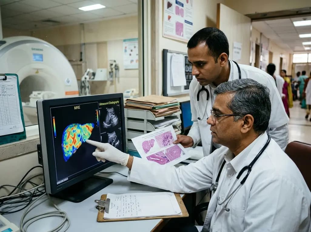
A comparative study reveals magnetic resonance elastography outperforms ultrasound elastography in assessing liver fibrosis in Fontan-associated liver disease, demonstrating stronger histological agreement and superior discrimination.
Today
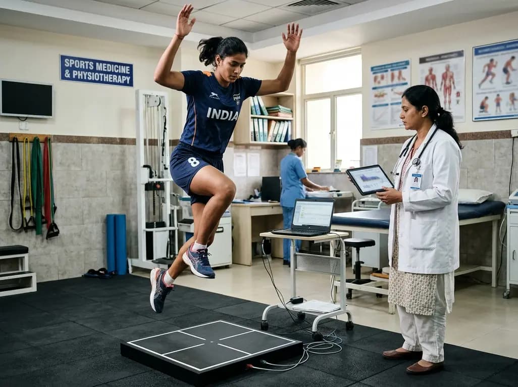
A systematic review and meta-analysis highlights the substantial impact of plyometric jump training on jump performance, reactive strength, and landing kinematics in female team-sport athletes, providing evidence-based programming guidelines for sports medicine and orthopedics.
Today

A comprehensive review of the role of growth hormone in diabetic kidney disease and podocytopathy. Learn how elevated GH activates ZEB2, Notch, and TGF-beta1, driving podocyte loss and glomerulosclerosis, and explore novel targeted therapeutic approaches.
Today
