.webp&w=384&q=75)

Loading, please wait...
Loading, please wait...
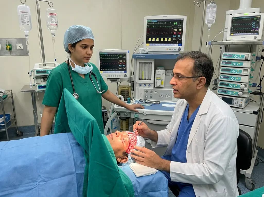
Awake craniotomy has emerged as a gold standard neurosurgical intervention for resecting intra-axial lesions located within or adjacent to eloquent brain regions. The primary goal of this procedure involves maximizing surgical resection margins while preserving critical motor, sensory, and language functions. However, maintaining physiological stability throughout the dynamic operative phases presents substantial clinical challenges for neuroanesthesiologists and surgical teams. Specifically, awake craniotomy hemodynamic management plays a vital role in sustaining adequate cerebral perfusion pressure, minimizing blood loss, and mitigating perioperative neurological deficits. Because surgical stimuli vary across pinning, craniotomy, cortical mapping, and tumor resection, hemodynamic fluctuations occur frequently. Consequently, clinicians must implement evidence-based neuroanesthetic strategies to prevent both acute hypertensive surges and severe hypotensive episodes. Systematic hemodynamic control directly influences patient cooperation during mapping tasks and ensures favorable long-term neurocognitive outcomes.
Intraoperative blood pressure variability remains a frequent complication during awake neurosurgical procedures. Clinical studies reveal that intraoperative hypertension occurs in approximately 8% to 34% of patients undergoing awake craniotomy. This hypertensive response typically arises during intensely stimulating surgical steps, such as head frame placement, scalp incision, and dural manipulation. Furthermore, emergence from sedation during the awake phase can precipitate intense anxiety and sympathetic activation. Significant risk factors for marked blood pressure elevation include previous craniotomy, pre-existing chronic hypertension, glioma pathology, and baseline impairment of cerebral autoregulation. Conversely, intraoperative hypotension affects approximately 10% to 11% of surgical candidates. Hypotension frequently stems from deep sedation, rapid postural adjustments, hypovolemia, or pharmacological side effects of infused anesthetics. Because brain tissue in peri-tumoral areas exhibits fragile microvasculature, extreme blood pressure swings disrupt localized cerebral homeostasis. Therefore, multidisciplinary surgical teams must identify patient-specific risk profiles preoperatively to anticipate and mitigate sudden hemodynamic variations before cortical mapping begins.
Hemodynamic instability during intracranial operations directly compromises cerebral perfusion pressure and microvascular tissue oxygenation. Evidence indicates that uncontrolled cerebral hypoperfusion significantly increases perioperative morbidity, with intraoperative stroke rates reaching up to 23% in vulnerable cohorts. In addition, persistent hypoperfusion contributes to postoperative neurological deficits in as many as 68% of affected individuals. When mean arterial pressure falls below the lower limit of cerebral autoregulation, peri-tumoral brain tissue experiences immediate ischemic stress. Furthermore, secondary brain swelling can rapidly complicate surgical exposure, impair neurofunctional testing, and elevate intracranial pressure. Conversely, severe hypertensive spikes increase the danger of intracranial hemorrhage, disrupted hemostasis, and acute cerebral hyperperfusion syndrome. These hemodynamic extremes severely impede real-time cortical and subcortical stimulation testing by causing patient agitation, confusion, or sudden neurological deterioration. Consequently, maintaining individualized blood pressure targets represents an indispensable prerequisite for successful surgery. Tailored hemodynamic goals facilitate safe tumor debulking while safeguarding critical neural pathways.
Successful awake craniotomy hemodynamic management relies upon structured multimodal anesthetic protocols that balance patient comfort with rapid neurological responsiveness. Comprehensive regional scalp nerve blocks provide the foundational defense against nociceptive stimulation during skull pinning, incision, and bone flap creation. By blocking sensory branches of the trigeminal and cervical nerves with long-acting local anesthetics, clinicians substantially suppress noxious autonomic reflexes. Moreover, conscious sedation protocols incorporating dexmedetomidine offer notable advantages over traditional opioid regimens. Dexmedetomidine provides effective dose-dependent sedation and mild analgesia without causing respiratory depression or blunting cortical mapping responses. When clinicians require adjunctive anxiolysis, carefully titrated low-dose midazolam provides supplemental comfort while maintaining cognitive interaction. When acute hypertension develops despite preventive measures, short-acting intravenous agents represent the standard of care. Specifically, esmolol and labetalol allow rapid, titratable blood pressure reduction without causing rebound cerebral vasodilation or elevating intracranial pressure. Similarly, clinicians utilize phenylephrine or noradrenaline infusions to treat transient hypotensive episodes expeditiously.
Continuous physiological monitoring is essential for optimizing cerebral oxygenation and sustaining organ perfusion during awake craniotomies. Invasive blood pressure monitoring via an arterial line provides beat-to-beat evaluation. This real-time tracking allows immediate detection of dangerous hemodynamic deviations. Furthermore, non-invasive optical modalities like near-infrared spectroscopy assess regional cerebral tissue oxygen saturation continuously. Near-infrared spectroscopy delivers immediate alerts regarding occult hypoperfusion, even when systemic blood pressure values appear within standard acceptable ranges. In addition, advanced systems calculate continuous cerebral autoregulation indices. These indices help neuroanesthesiologists delineate individualized blood pressure thresholds for each patient. By understanding precise autoregulatory boundaries, the surgical team avoids empirical targets that might cause focal ischemia. Integrating electrophysiological monitoring alongside hemodynamic tracking ensures aggressive cytoreduction without compromising functional cortical networks.
Despite compelling clinical evidence supporting individualized hemodynamic strategies, significant practice variation currently persists across international neurosurgical centers. A prominent contributing factor is the paucity of large-scale randomized controlled clinical trials comparing distinct neuroanesthetic protocols for awake craniotomies. Consequently, establishing standardized institutional guidelines represents an urgent priority for modern neuroanesthesia societies. Looking ahead, technological innovations and digital health tools will transform intraoperative hemodynamic and neuroprotective care. Machine learning models and real-time artificial intelligence algorithms can predict hypotension and hypertensive surges minutes before clinical manifestation. Predictive algorithms analyze multi-parameter waveforms from arterial lines and optical sensors to empower preemptive clinical interventions. Furthermore, closed-loop automated target-controlled infusion systems may soon regulate anesthetic depth and vasoactive infusions simultaneously. Ultimately, combining artificial intelligence with comprehensive neuromonitoring will refine cerebral protection. This integrated approach maximizes tumor resection and safeguards patient safety.
Intraoperative hypertension occurs in up to 34% of patients undergoing awake craniotomy, primarily triggered by nociceptive stimulation from skull pinning, dural manipulation, or surgical incision. Additionally, emergence from deep sedation into the awake state often induces substantial anxiety, distress, and sympathetic activation. Pre-existing chronic hypertension, recurrent craniotomy procedures, and tumor-induced disruption of local cerebral autoregulation further heighten the risk of severe intraoperative blood pressure spikes during functional mapping.
Intraoperative hypotension substantially reduces cerebral perfusion pressure, particularly within vulnerable peri-tumoral brain tissues that lack intact autoregulatory capacity. Even brief hypotensive episodes correlate strongly with perioperative ischemic stroke, which occurs in up to 23% of compromised cases. Consequently, inadequate cerebral microvascular blood flow induces tissue hypoxia, exacerbates postoperative neurological deficits in up to 68% of patients, and increases overall perioperative morbidity and mortality rates significantly.
Neuroanesthesiologists favor highly titratable, short-acting pharmacological agents to maintain strict hemodynamic stability without compromising cortical mapping. Continuous dexmedetomidine infusions provide excellent conscious sedation and analgesia without inducing respiratory depression. When acute hypertensive surges occur, selective beta-blockers such as esmolol and combined alpha-beta antagonists like labetalol rapidly normalize blood pressure without increasing intracranial pressure. Conversely, titrated phenylephrine or noradrenaline infusions promptly correct unexpected intraoperative hypotension.
Disclaimer: This content is for informational and educational purposes only... Refer to the latest local and national guidelines for clinical practice.
References

Read summarized clinical updates, watch expert medical content, and earn CME certifications right from your smartphone.
Open your smartphone camera to scan and install.





Intraoperative hemodynamic stability during awake craniotomy is essential to prevent cerebral hypoperfusion and stroke. This review explores evidence-based multimodal strategies, individualized blood pressure targets, advanced neuromonitoring, and emerging AI tools for optimal cerebral protection.
4 weeks back

A comprehensive systematic review reveals that CT-based epicardial adipose tissue radiomics combined with machine learning holds significant promise for predicting atrial fibrillation occurrence and recurrence. However, technical heterogeneity and validation gaps currently limit immediate clinical adoption.
Today
A retrospective cohort study evaluated 1,000 low-risk singleton births to assess maternal and neonatal outcomes during office-hours versus off-hours. Off-hours deliveries were significantly associated with higher odds of intrapartum cesarean sections and episiotomies, highlighting critical obstetric staffing considerations.
Today
Procedural success in panvascular intervention does not guarantee vessel stability. The tri-ecological framework introduces vascular device suitcordance and the Suitcordance Score, matching mechanical, cellular, and physicochemical-immune dynamics to restore true vessel homeostasis and prevent late failures.
Today

Researchers have engineered an artificial sensory-pain integrated receptor using a multi-threshold organic synaptic transistor. The system distinguishes benign touch from noxious stimuli, enabling safer, more responsive prosthetic limbs with built-in memory and simplified hardware architecture.
Today

Medical robotics is transitioning beyond operative suites into diagnostics, cardiology, and endocrinology. Explore how innovations in tele-robotics, AI integration, and nanomedicine are overcoming geographical barriers and transforming precision healthcare delivery across India's evolving disease landscape.
Today
