.webp&w=384&q=75)

Loading, please wait...
Loading, please wait...

Autoimmune encephalitis represents a severe group of neuroinflammatory disorders that cause subacute cognitive decline, refractory seizures, and psychiatric manifestations. Specifically, anti-leucine-rich glioma inactivated-1 and anti-contactin-associated-protein-2 disorders constitute two prevalent subtypes. Although pathogenic autoantibodies targeting cell-surface antigens drive these conditions, the cellular orchestration governing antibody production has remained elusive. Consequently, a landmark multi-omic investigation now uncovers crucial mechanistic insights. The study demonstrates that coordinated T-cell activation and innate-like lymphocyte deficiencies directly promote central humoral autoimmunity.
Clinicians frequently encounter a puzzling diagnostic paradox when evaluating patients with suspected limbic encephalitis. Standard cerebrospinal fluid analysis often displays completely normal leukocyte counts, normal glucose, and unremarkable total protein concentrations. However, high-resolution single-cell RNA sequencing reveals intense, localized humoral activity within the central nervous system. Researchers observed profound clonal expansion of antibody-secreting cells exclusively within patient cerebrospinal fluid. In contrast, parallel peripheral blood samples exhibited far lower frequencies of these specialized cells. Furthermore, detailed transcriptional profiling identified these antibody-secreting cells predominantly as active plasmablasts. These cells transcribed immunoglobulin heavy chains belonging to the IgG4 and IgG1 or IgG2 subclasses. Moreover, bioinformatic analysis confirmed extensive somatic hypermutation, providing undeniable evidence of local affinity maturation. Recombinant human monoclonal antibodies generated from these expanded clones bound specifically and robustly to native LGI1 or CASPR2 autoantigens. Therefore, even when routine cytological examination suggests an absence of inflammation, the cerebrospinal compartment harbors an active, highly specialized antigen-driven plasmablast niche. These findings establish that normal baseline fluid cytology does not exclude aggressive intrathecal B-cell activation.
While autoantibodies directly damage synaptic machinery, pathogenic B cells require critical co-stimulatory assistance to sustain high-affinity antibody secretion. The investigation revealed that both CD4-positive and CD8-positive T-cell compartments undergo marked shifts during acute disease. Specifically, central memory T cells demonstrated prominent activation markers alongside significant clonal restriction. Rather than presenting a broad, nonspecific inflammatory response, these alpha-beta T cells displayed selective clonal expansion within the cerebrospinal fluid. Additionally, researchers identified matching T-cell receptor clones shared between cerebrospinal fluid and peripheral blood compartments. This crucial finding indicates active bidirectional trafficking across the blood-brain barrier. CD4 central memory helper cells likely orchestrate germinal center-like responses within neural niches, thereby driving plasmablast differentiation. Meanwhile, clonally expanded CD8 central memory cells could mediate localized cytotoxic signaling or release pro-inflammatory cytokines that exacerbate blood-brain barrier permeability. Consequently, these synchronized alpha-beta T-cell responses provide the indispensable scaffolding required for enduring autoantibody production. Recognizing this critical T-cell involvement alters our understanding of disease pathophysiology, shifting the clinical paradigm from a purely humoral view toward a coordinated neuro-immune axis.
Beyond conventional alpha-beta T cells, innate-like lymphocytes play a decisive role in maintaining systemic and tissue-level immune tolerance. The study highlighted a striking and consistent reduction of mucosa-associated invariant T cells. Patients exhibited profound MAIT cell depletion within the cerebrospinal fluid in LGI1 encephalitis and across peripheral blood in both disease subtypes. Furthermore, flow cytometric analysis in independent patient cohorts validated this marked numerical drop. Concurrently, investigators observed distinct compositional alterations among natural killer cell subsets, reflecting extensive disruption of innate cellular networks. Under physiological conditions, MAIT cells express semi-invariant T-cell receptors that recognize microbial metabolites presented by MR1 molecules. Moreover, these unique lymphocytes secrete regulatory cytokines that actively suppress aberrant autoantibody generation. When mucosal or peripheral regulatory checks falter, autoreactive B cells escape standard suppressive checkpoints. Consequently, the severe depletion of circulating and central MAIT cells removes a vital homeostatic brake. This loss permits unchecked clonal expansion of autoreactive plasmablasts, highlighting innate immunodeficiency as an unexpected driver of central nervous system autoimmunity.
To determine whether observed cellular alterations reflect true neuropathology rather than peripheral epiphenomena, investigators analyzed autopsied brain specimens from affected individuals. Histopathological examination confirmed that MAIT-like T cells physically infiltrate inflamed parenchymal brain tissue in both LGI1 and CASPR2 encephalitis. Thus, innate-like lymphocytes migrate directly to damaged brain regions during active neuroinflammation. Subsequently, researchers tested the functional consequences of MAIT cell deficiency using an in vivo immunization paradigm. Genetically modified mice lacking MAIT cells received active immunization against LGI1 and CASPR2 autoantigens. Remarkably, MAIT-deficient animals exhibited significantly higher rates of seroconversion compared to wild-type controls. In addition, these knockout animals developed substantially elevated serum autoantibody titers. Therefore, experimental data confirm that functional MAIT cells exert a powerful suppressive effect against antineuronal antibody production. When innate regulatory surveillance declines, the immune system fails to control autoreactive B-cell lineages. Together, histopathological detection and preclinical knockout assays conclusively demonstrate that mucosal and invariant T cells serve as critical guardians against pathogenic neuro-autoimmunity.
These comprehensive discoveries carry profound implications for practicing neurologists and critical care specialists managing acute encephalopathy. First, clinicians must remember that normal routine cerebrospinal fluid findings never rule out autoimmune encephalitis. Because pathogenic plasmablasts expand intrathecally despite normal cell counts, clinicians must maintain a low threshold for ordering dedicated neural autoantibody panels. Delayed diagnosis often leads to irreversible hippocampal atrophy and chronic cognitive morbidity. Second, the confirmation of active central memory T-cell expansion challenges conventional treatment algorithms. Standard first-line therapies such as intravenous corticosteroids, plasma exchange, and intravenous immunoglobulins offer rapid symptomatic control. However, refractory or relapsing cases often necessitate second-line agents. Clinicians frequently prescribe rituximab to deplete CD20-positive B cells. Nevertheless, rituximab spares long-lived plasma cells and mature plasmablasts. Furthermore, rituximab does not directly suppress pathogenic memory T cells. Given the prominent involvement of clonally expanded CD4 and CD8 T cells, therapeutic regimens targeting both T-cell co-stimulation and plasma cell longevity could substantially improve durable clinical remissions. Consequently, combined immunomodulation represents the next rational frontier in patient management.
Routine cerebrospinal fluid analysis frequently reveals normal cell counts and protein levels in LGI1 and CASPR2 encephalitis. Consequently, clinicians may mistakenly rule out active central neuroinflammation. Advanced molecular diagnostics demonstrate that pathogenic antibody-secreting plasmablasts expand intrathecally despite unremarkable routine cytology, necessitating specialized neural antibody testing.
Central memory CD4 and CD8 T cells undergo clonal expansion and activation within the cerebrospinal fluid. Furthermore, these cells circulate between peripheral blood and brain compartments. They provide essential co-stimulatory support to autoreactive B cells and directly amplify localized neuroinflammatory cascades, driving disease progression and long-term neurocognitive impairment.
Mucosa-associated invariant T cells act as key immune regulators that restrain pathogenic humoral activity. However, patients experience marked MAIT cell depletion in both blood and cerebrospinal fluid. Preclinical models confirm that MAIT cell deficiency accelerates autoantibody production and elevates serum titers, indicating that failure of innate immune suppression accelerates disease onset.
Disclaimer: This content is for informational and educational purposes only... Refer to the latest local and national guidelines for clinical practice.
References

Read summarized clinical updates, watch expert medical content, and earn CME certifications right from your smartphone.
Open your smartphone camera to scan and install.





A breakthrough study in Brain reveals that LGI1 and CASPR2 autoimmune encephalitis feature intrathecal clonal expansion of plasmablasts, activated central memory αβ T cells, and a striking loss of regulatory MAIT cells, reshaping our understanding of neuroinflammation and guiding targeted immunotherapy.
Today
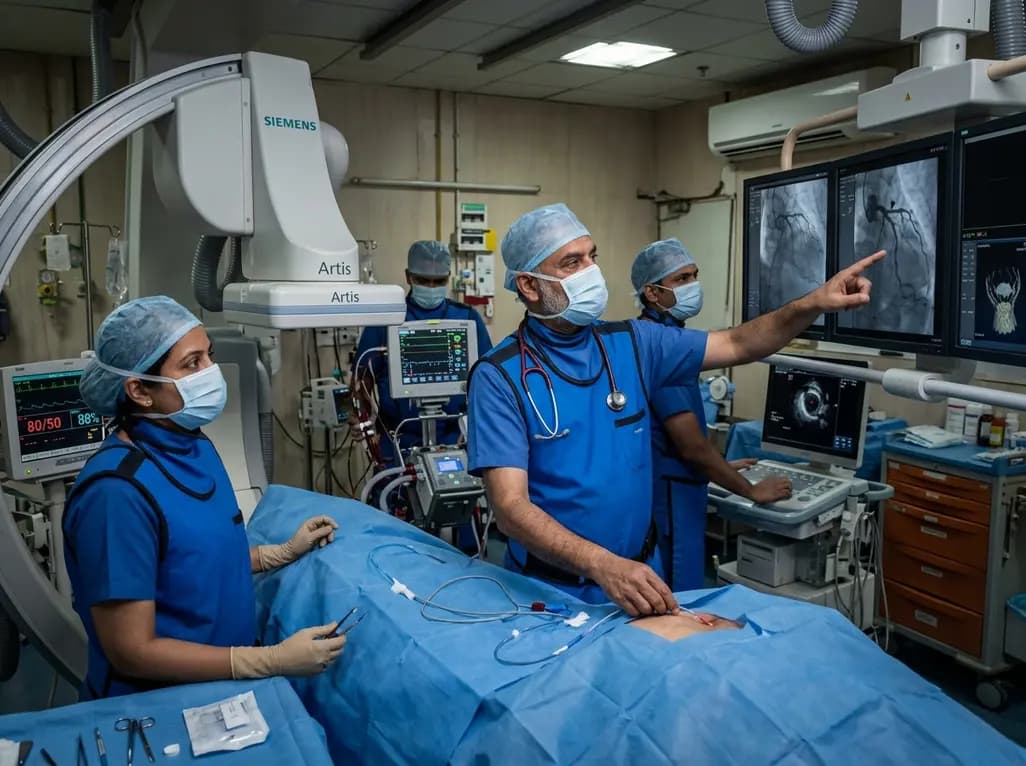
A rare case of acute coronary obstruction during TAVR caused by suspected calcified amorphous tumor embolization underscores the critical role of intracardiac imaging, emergency ECMO, and the paradox of enlarged-aperture valve geometry.
Today

Prelacteal feeding persists in South Asian communities despite known clinical risks. This review examines determinants from Chitral, pathophysiological hazards, and pediatric strategies to eliminate harmful feeds and support exclusive breastfeeding.
Today

Transcatheter mitral valve replacement in patients with degenerated bioprostheses carries a substantial risk of LVOT obstruction. Combining leaflet modification via the BATMAN technique with mechanical circulatory support offers a viable, life-saving solution for anatomically challenging surgical candidates.
Today

A comparative cohort study demonstrates that patients undergoing total hip arthroplasty after prior hip arthroscopy achieve comparable functional recovery, low complication rates, and equivalent implant survivorship when conversion occurs after six months.
Today

A cross-sectional study of 320 adults evaluated the link between habitual fasting duration and dietary quality using Nutrition Quotient scores, highlighting the clinical necessity of balancing time-restricted eating with nutrient density and meal quality in routine metabolic practice.
Today
