

Loading, please wait...
Loading, please wait...
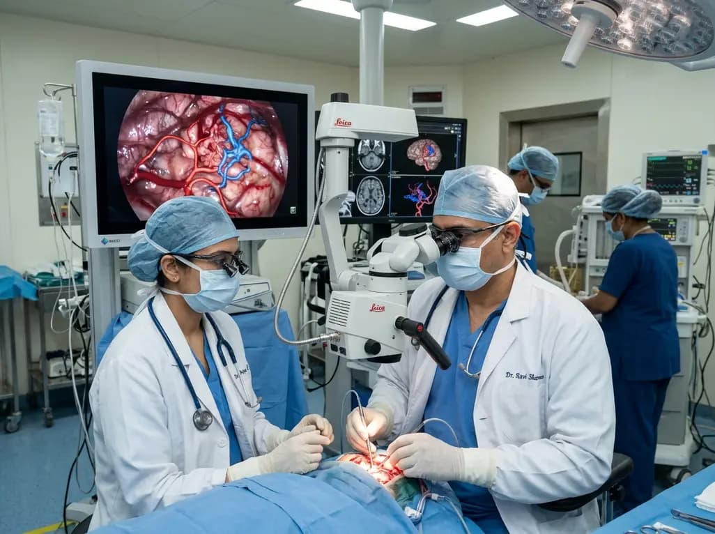
Microsurgical excision remains a definitive curative treatment for intracranial vascular lesions, but brain AVM resection presents distinct technical hurdles. Early proximal arterial control constitutes an established surgical principle, yet deep or obscured feeding vessels often compromise straightforward exposure. When surgeons encounter hidden pedicles beneath cerebral convolutions, dissecting the nidus prematurely can provoke life-threatening hemorrhage. Consequently, neurosurgeons have sought image-guided workflows that reveal complex angioarchitecture prior to parenchymal incision. In a landmark longitudinal study of 158 consecutive patients, researchers evaluated an integrated workflow combining microscope-based augmented reality with multimodal neuronavigation. By translating sophisticated preoperative vascular datasets into actionable operative targets, this protocol significantly improves operative precision. Ultimately, this paradigm shift allows surgical teams to systematically eliminate high-pressure arterial feeders while protecting critical surrounding brain parenchyma.
Successful execution of this approach begins with comprehensive preoperative imaging. Specifically, clinicians assemble case-specific datasets incorporating computed tomography angiography, high-resolution structural magnetic resonance imaging, magnetic resonance angiography, and three-dimensional rotational digital subtraction angiography. Standard clinical neuronavigation platforms fuse these disparate imaging modalities into a single cohesive anatomical volume. Next, the surgical team meticulously segments all angiographically identifiable arterial feeders along their surgically relevant trajectories. In addition, the neurosurgeon prioritizes these vessels as discrete sequential targets during craniotomy and corridor planning. For lesions situated within or adjacent to eloquent cortex, the protocol selectively coregisters functional magnetic resonance imaging and diffusion tensor imaging tractography. Therefore, the operating team obtains a clear understanding of critical white matter pathways before entering the operating suite. This detailed structural roadmap reduces anatomical ambiguity and helps clinicians select the safest trajectory toward deep vascular pedicles.
During microsurgical exploration, surgeons display the segmented vascular targets directly within the operative visual field using microscope-based augmented reality. Furthermore, this dynamic heads-up display projects color-coded arterial feeders onto the optical path, aligning virtual models with actual brain anatomy. As a result, the operating neurosurgeon can pinpoint deep feeder trajectories without constantly looking away toward external navigation monitors. This continuous visualization proves exceptionally beneficial when feeding vessels lie buried within deep sulci or beneath overlying arterialized veins. Moreover, the surgeon sequentially clips and divides the arterial supply prior to undertaking substantial nidal dissection. Halting high-flow arterial inputs early in the operation substantially decompresses the malformation. Consequently, the nidus softens and shrinks, which markedly minimizes intraoperative bleeding and facilitates subsequent circumferential dissection. By turning complex three-dimensional angioarchitecture into an orderly checklist of operative targets, the workflow transforms an unpredictable procedure into a disciplined sequence.
The longitudinal clinical trial evaluated 158 consecutive navigation-assisted resections across a broad spectrum of Spetzler-Martin grades I through V. Remarkably, complete malformation obliteration on initial postoperative angiography reached 97.5%, representing 154 of the 158 surgical cases. In addition, long-term functional results demonstrated outstanding neurological preservation across diverse patient cohorts. At a median follow-up of 15 months, 93.0% of treated individuals maintained functional independence, achieving modified Rankin Scale scores between 0 and 2. Although an increase in modified Rankin Scale score occurred in 18.4% of patients, these changes most commonly reflected minor, nondisabling neurological deficits. Importantly, permanent loss of functional independence directly attributable to surgical intervention occurred in only 3.1% of patients, while the overall mortality rate remained low at 1.3%. Thus, the structured feeder-directed strategy delivers exceptional angiographic cure rates while maintaining a favorable morbidity profile even in complex, high-grade cerebrovascular malformations.
Early proximal arterial control addresses one of the most perilous hazards in vascular neurosurgery: normal perfusion pressure breakthrough and catastrophic intraoperative hemorrhage. When surgeons disturb venous outflow before arresting incoming arterial feeders, high intravascular pressure within the nidus causes sudden, massive extravasation. However, augmented reality guidance allows the surgeon to systematically eliminate high-pressure arterial inputs while preserving essential draining veins until final excision. Consequently, intravascular pressure within the vascular nidus plummets before circumferential parenchymal dissection begins. Furthermore, integrating tractographic datasets helps avoid unnecessary mechanical traction on surrounding corticospinal and optic radiations. This protective measure prevents secondary ischemic injury in fragile perilesional brain parenchyma. In addition, reducing reliance on extensive preoperative endovascular embolization may protect patients from embolization-related ischemic strokes and vessel perforations. Therefore, early feeder identification significantly enhances overall procedural safety and improves microsurgical predictability.
Beyond individual patient outcomes, this integrated multimodal workflow provides immense pedagogical value for cerebrovascular training programs. Historically, mastering brain arteriovenous malformation resection required decades of clinical experience to conceptualize complex vascular volumes mentally. In contrast, microscope-based augmented reality creates an intuitive visual bridge between two-dimensional angiographic findings and three-dimensional surgical topography. Trainees and junior neurosurgeons can observe exactly how deep feeding pedicles branch from main arterial trunks beneath the cortical surface. Additionally, surgical teams can simulate various dissection corridors and approach trajectories during preoperative multidisciplinary conferences. Modern neurosurgical suites in tertiary hospitals can readily integrate these navigation protocols using existing neuronavigation hardware and commercial microscope interfaces. As software algorithms and robotic visualization platforms continue to evolve, real-time spatial guidance will become an indispensable standard of care. Ultimately, adopting this systematic approach empowers surgical teams to tackle formidable cerebrovascular lesions with greater confidence and reproducible clinical success.
Microscope-based augmented reality overlays segmented vascular models directly onto the surgical field, enabling neurosurgeons to visualize hidden arterial feeders in real time. Consequently, surgeons identify and control high-pressure pedicles before nidal dissection without diverting their gaze from the operating microscope. This seamless visual integration enhances spatial orientation, shortens operative duration, minimizes blood loss, and helps avoid accidental damage to adjacent eloquent brain parenchyma or critical venous outflow pathways.
Early proximal arterial occlusion significantly reduces intravascular pressure throughout the malformation nidus before circumferential dissection begins. As a result, the vascular nidus collapses and softens, making surgical manipulation far safer and substantially reducing intraoperative hemorrhage. Furthermore, preserving draining veins while systematically eliminating arterial feeding vessels prevents acute venous hypertension and reduces the risk of normal perfusion pressure breakthrough swelling in adjacent cerebral tissues.
Although augmented reality provides precise anatomical guidance, it does not replace intraoperative or postoperative digital subtraction angiography. Augmented reality displays static preoperative vascular datasets, whereas angiography evaluates real-time hemodynamic flow and verifies immediate lesion obliteration. Therefore, neurosurgeons combine augmented reality feeder targeting with intraoperative indocyanine green videoangiography or conventional angiography to ensure complete nidus exclusion and confirm the preservation of normal passage vessels.
Disclaimer: This content is for informational and educational purposes only... Refer to the latest local and national guidelines for clinical practice.
References

Read summarized clinical updates, watch expert medical content, and earn CME certifications right from your smartphone.
Open your smartphone camera to scan and install.





A 158-case longitudinal study demonstrates that integrating microscope-based augmented reality with multimodal navigation enables precise sequential arterial feeder targeting during brain AVM resection, achieving 97.5% complete obliteration and preserving functional independence in 93% of patients.
Today

Delhi civic authorities have cleared 1,066 contractual public health posts and a Rs 923.6-crore road-sweeping initiative to tackle vector-borne illnesses and air pollution. Learn how enhanced entomological surveillance, anti-larval drives, and dust control bolster community preventive medicine and clinical outcomes.
Today

Medical experts caution that GLP-1 receptor agonists offer metabolic support rather than a curative answer for polyendocrine metabolic ovarian syndrome. Clinicians must evaluate glycemic markers, reproductive plans, and lifestyle interventions while discouraging unsupervised medication use among female patients.
Today

A retrospective CAHAL study comparing Amplatzer and Occlutech devices in 413 patients found no significant difference in moderate-to-large residual shunt rates at 6 months follow-up after percutaneous PFO closure, supporting clinical equivalence between both platforms.
Today

Age-related spinal kyphosis is driven by vertebral fragility, disc degeneration, and paraspinal muscle dysfunction. Coexisting with locomotive syndrome and frailty, its management requires combined anti-osteoporotic therapy, spinal extensor rehabilitation, and comprehensive nutritional optimization.
Today

A longitudinal feasibility pilot study highlights that combining polygenic risk score disclosure with in-person lifestyle counseling significantly enhances genomic literacy in individuals at risk for type 2 diabetes, overcoming educational disparities and reinforcing patient motivation.
Today
