.webp&w=384&q=75)

Loading, please wait...
Loading, please wait...
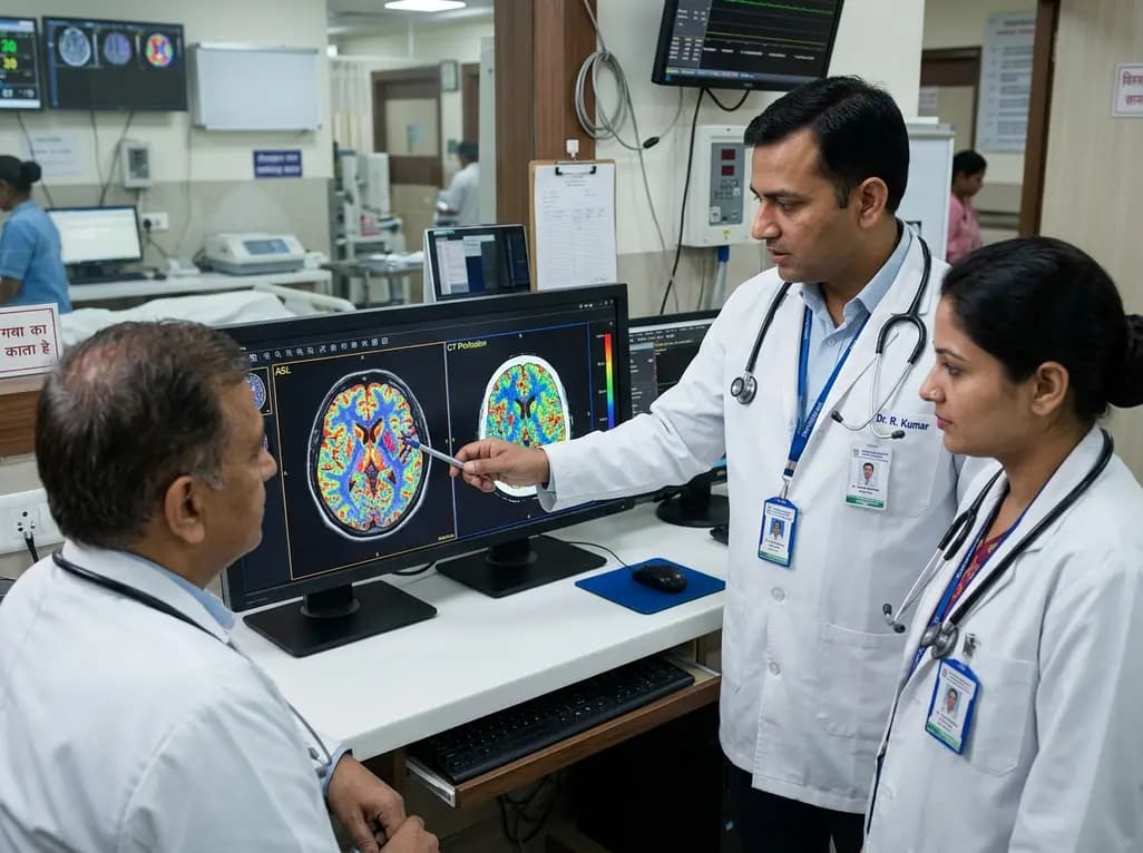
Moyamoya disease represents a progressive, steno-occlusive intracranial vasculopathy characterized by stenosis of the terminal internal carotid arteries and compensatory collateral vessel formation. Precise evaluation of cerebral hemodynamics is critical for establishing disease stage, determining stroke risk, and planning revascularization surgery. The routine application of ASL in moyamoya angiopathy has gained substantial clinical interest because clinicians require accurate, repeatable, and non-invasive methods to assess perfusion. Traditionally, gold-standard modalities like positron emission tomography require radioactive tracers and specialized facilities that remain inaccessible to most centers. Although computed tomography perfusion provides rapid quantification, it carries risks of ionizing radiation and iodinated contrast nephropathy. Consequently, arterial spin labeling with acetazolamide challenge has emerged as an attractive diagnostic tool. Recent retrospective evidence shows that this magnetic resonance technique matches computed tomography perfusion in predicting key angiographic outcomes.
Cerebrovascular reserve reflects the physiological capacity of cerebral arterioles to dilate in response to vasodilatory stimuli. In chronic ischemic disorders such as moyamoya angiopathy, primary basal vessels suffer progressive narrowing. As a result, downstream autoregulatory microvessels maximally dilate even under resting baseline conditions to preserve adequate cerebral blood flow. When metabolic demand rises or perfusion pressure drops further, these vessels cannot dilate additionally, leaving brain tissue vulnerable to profound ischemia. Therefore, quantifying cerebrovascular reserve provides essential insight beyond baseline resting perfusion. Clinicians commonly use acetazolamide, a carbonic anhydrase inhibitor, to induce transient maximal arteriolar vasodilation. Regions with exhausted autoregulation fail to augment local blood flow after acetazolamide administration. In severe cases, blood flow paradoxically diverts away from chronically dilated territories toward healthy vascular beds, creating an intracerebral steal phenomenon. Identifying these vulnerable zones enables timely surgical revascularization before irreversible cerebral infarction occurs.
Arterial spin labeling utilizes endogenous blood water protons as a freely diffusible tracer, eliminating any requirement for exogenous gadolinium contrast. During pseudo-continuous arterial spin labeling, radiofrequency pulses magnetically label arterial blood in cervical vessels before it travels into the intracranial capillary bed. Subtracting labeled images from control images produces quantitative cerebral blood flow maps. When investigators pair baseline arterial spin labeling with post-acetazolamide acquisition, they calculate relative cerebrovascular reserve as the percentage change in cerebral blood flow across specific territories. Furthermore, multi-delay and pseudo-continuous protocols help mitigate transit delay artifacts that typically arise from sluggish collateral flow in stenotic cerebral vasculature. Because the technique involves neither ionizing radiation nor nephrotoxic agents, clinicians can safely perform repeated assessments across longitudinal follow-ups. Consequently, utilizing ASL in moyamoya angiopathy offers a comprehensive physiological evaluation that reliably mirrors complex intracranial hemodynamic rearrangements without compromising patient safety.
Digital subtraction angiography remains the reference standard for morphological staging via the classic Suzuki classification system and for quantifying proximal intracranial stenosis. However, catheter angiography carries procedural risks and does not measure tissue-level perfusion directly. A recent comparative investigation analyzed thirty-nine patients with moyamoya angiopathy to assess how arterial spin labeling metrics correlate with angiographic severity. Territory-specific cerebral blood flow was extracted across the anterior, middle, and posterior cerebral artery distributions. Logistic regression models combining baseline cerebral blood flow, acetazolamide-derived cerebrovascular reserve, and patient age demonstrated moderate to good discriminatory ability for advanced Suzuki stages and severe proximal A1 or M1 segment arterial stenosis. Specifically, receiver operating characteristic analyses yielded area under the curve values ranging between 0.71 and 0.85 across these anatomical endpoints. These findings confirm that non-invasive physiological parameters closely parallel macrovascular structural narrowing.
Historically, computed tomography perfusion has served as a widely available modality for rapid cerebrovascular evaluation. However, comparative analyses reveal no statistically significant differences in diagnostic discrimination between arterial spin labeling and computed tomography perfusion models. Across shared endpoints, computed tomography perfusion models achieved area under the curve values ranging from 0.69 to 0.74, closely matching the performance of arterial spin labeling. Most notably, arterial spin labeling models achieved their highest predictive accuracy when forecasting postoperative collateral status, reaching an area under the curve of 0.85. Computed tomography perfusion inevitably exposes patients to radiation and requires iodinated contrast agents that carry risks of allergic reactions and renal toxicity. Because moyamoya angiopathy predominantly affects children and young adults requiring repeated lifetime imaging, eliminating radiation exposure is a paramount clinical objective. Thus, arterial spin labeling provides an equivalent diagnostic yield while markedly improving long-term safety profiles.
Extracranial-to-intracranial bypass procedures, including direct superficial temporal artery to middle cerebral artery anastomosis and indirect encephaloduroarteriosynangiosis, aim to restore adequate regional perfusion and prevent future ischemic events. Preoperative identification of exhausted cerebrovascular reserve serves as a primary indication for revascularization. Postoperatively, evaluating neovascularization and surgical bypass patency is critical for determining long-term procedural success. Arterial spin labeling derived metrics reliably predict postoperative collateral development, assisting neurosurgeons in monitoring graft maturation and regional hemodynamic recovery over time. In addition, post-surgical arterial spin labeling scans can detect transient focal hyperperfusion, a well-recognized complication following direct bypass that presents with neurological deficits or intracerebral hemorrhage. Early detection of regional hyperperfusion allows timely blood pressure regulation and neuroprotective management. Therefore, incorporating non-invasive perfusion magnetic resonance imaging into standardized perioperative workflows optimizes both surgical decision-making and patient outcomes.
Although acetazolamide-challenged arterial spin labeling demonstrates notable diagnostic parity with traditional perfusion imaging, clinicians must interpret quantitative findings within specific clinical contexts. First, severe arterial transit delays common in advanced moyamoya disease can occasionally cause underestimation of true cerebral blood flow if label arrival times exceed the chosen post-labeling delay. Adopting multi-delay pseudo-continuous protocols effectively resolves these transit artifacts. Second, intravenous acetazolamide administration occasionally induces minor side effects such as transient perioral numbness, dizziness, headache, or mild nausea, requiring careful pre-procedure screening and patient monitoring. Contraindications including known sulfonamide allergies, severe hepatic dysfunction, or severe renal impairment require strict pre-scan evaluation. Despite these technical nuances, implementing standardized non-contrast perfusion MRI protocols significantly improves neurovascular diagnostic workflows. Future prospective multicenter trials will further refine quantitative thresholds, solidifying non-invasive hemodynamic imaging as a frontline standard of care.
Arterial spin labeling does not require ionizing radiation or iodinated contrast media, making it completely non-invasive and safe for serial monitoring. It provides quantitative cerebral blood flow and cerebrovascular reserve measurements comparable in accuracy to CT perfusion without exposing young patients to cumulative radiation or nephrotoxicity risks.
Acetazolamide induces potent intracranial arteriolar vasodilation by inhibiting carbonic anhydrase. Healthy vascular beds exhibit significant increases in cerebral blood flow post-administration. Conversely, chronically ischemic territories with pre-existing maximal compensatory vasodilation show minimal or no blood flow augmentation, highlighting exhausted cerebrovascular reserve or regional steal phenomena.
Yes, preoperative and postoperative arterial spin labeling models demonstrate strong predictive performance for evaluating collateral status and bypass maturation, achieving area under the curve values up to 0.85. Furthermore, postoperative scans help clinicians identify regional hyperperfusion syndrome early, allowing prompt blood pressure management to prevent complications.
Disclaimer: This content is for informational and educational purposes only and should not be taken as professional medical advice. Always consult a qualified healthcare provider for diagnosis and treatment decisions. Refer to the latest local and national guidelines for clinical practice.
References
Serdyuk V et al. Acetazolamide-challenged ASL shows comparable performance to CT perfusion for angiographic outcomes in moyamoya angiopathy. Clin Neurol Neurosurg. 2026 Sep. doi: 10.1016/j.clineuro.2026.109517. PMID: 42190491.
Federau C, Christensen S, Zun Z, et al. Cerebral blood flow, transit time, and apparent diffusion coefficient in moyamoya disease before and after acetazolamide. Neuroradiology. 2017;59(1):5-12.
Wang R, Liu X, Xiao W, et al. Monitoring Cerebral Perfusion Changes after Revascularization in Patients with Moyamoya Disease by Using Arterial Spin-labeling MR Imaging. Radiology. 2018;289(1):175-182.

Read summarized clinical updates, watch expert medical content, and earn CME certifications right from your smartphone.
Open your smartphone camera to scan and install.





A recent study demonstrates that acetazolamide-challenged arterial spin labeling (ASL) MRI achieves comparable predictive performance to CT perfusion for evaluating angiographic severity and postsurgical collateral status in moyamoya angiopathy, offering a safe, non-invasive imaging alternative.
4 weeks back
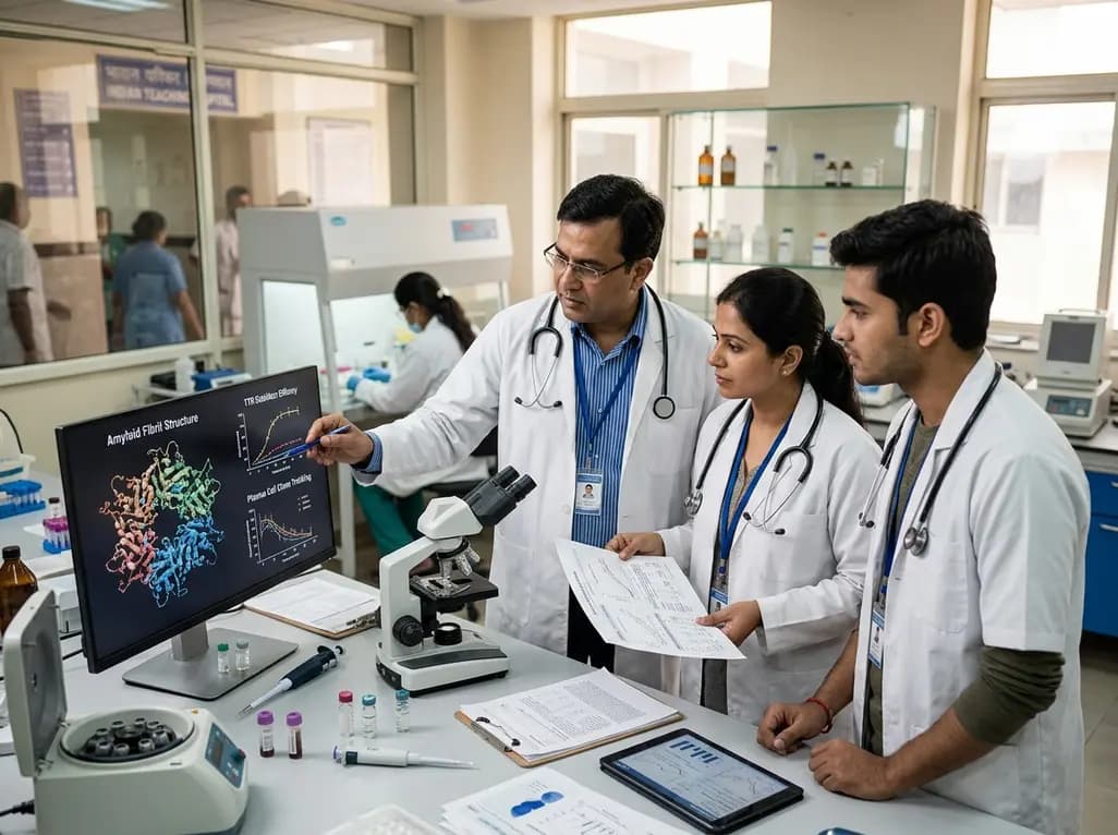
Systemic amyloidosis management is undergoing a therapeutic revolution. This clinical review synthesizes updates from the International Society of Amyloidosis, detailing novel plasma cell therapies, TTR stabilizers, silencers, translational models, and artificial intelligence in drug development.
Today
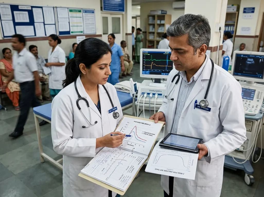
A retrospective study of 824 patients undergoing over 2,200 intradetrusor onabotulinumtoxinA injections identifies sex- and etiology-specific predictors of 2-week post-void residual, highlighting the influence of baseline residual volumes, toxin dosage, and distinct urodynamic parameters on urinary retention risk.
Today
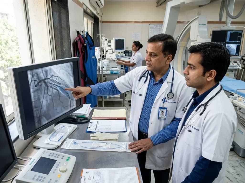
The US FDA has granted approval to the SELUTION SLR drug-eluting balloon for treating coronary in-stent restenosis. This milestone introduces a sustained-release sirolimus platform that eliminates the need for additional permanent metallic stents while maintaining robust vascular healing and luminal patency.
Today

Posttraumatic pisotriquetral instability is a rare wrist injury causing severe hypothenar pain and weakness. Explore clinical examination, imaging, and pisiformectomy.
Today

A comprehensive review analyzes periconceptional GLP-1 receptor agonist exposure, revealing reassuring data on congenital anomalies and adverse perinatal outcomes while emphasizing the need for cautious patient counseling.
Today
