.webp&w=384&q=75)

Loading, please wait...
Loading, please wait...

Forensic pathologists often encounter substantial hurdles when determining the exact cause of death in submersion victims. Consequently, establishing an objective post-mortem diagnosis requires rigorous exclusion of alternative etiologies. Drowning typically leaves non-specific findings, such as pulmonary emphysema, fluid accumulation, and frothy airway secretions. However, these physical markers rapidly deteriorate during advanced decomposition. Therefore, forensic investigators actively seek dependable immunohistochemical tools to corroborate vital submersion. Recent scientific literature highlights the diagnostic value of cerebral aquaporin expression in drowning cases. Specifically, aquaporins serve as specialized water channel proteins that govern fluid movement across cerebral membranes. In addition, these channel proteins respond dynamically to osmotic changes during asphyxial submersion. As water enters the alveolar spaces, substantial hemodynamic and electrolyte shifts occur throughout the systemic circulation. These physiological alterations directly influence cerebral fluid transport mechanisms. Furthermore, researchers strive to distinguish antemortem submersion from post-mortem body disposal in water bodies. Traditional techniques, such as the diatom test, frequently yield ambiguous or conflicting results. Therefore, forensic experts require precise molecular targets within preserved parenchymal tissues. Glial water channels provide exceptional stability and diagnostic promise in such demanding medicolegal evaluations.
Different aquatic environments trigger distinct pathophysiological mechanisms during terminal submersion. Specifically, freshwater drowning involves the rapid aspiration of markedly hypotonic fluid into the pulmonary alveoli. Consequently, this hypotonic influx drives rapid water absorption across the alveolar-capillary barrier into the bloodstream. This sudden fluid movement causes profound hemodilution, severe intravascular hemolysis, and dangerous hyponatremia. Furthermore, the circulating hypotonic plasma generates a steep osmotic gradient across the blood-brain barrier. As a result, massive amounts of fluid enter cerebral astrocytes, precipitating acute cytotoxic edema. In contrast, saltwater submersion initiates an entirely opposite sequence of osmotic and physiological events. Seawater presents a distinctly hypertonic environment compared to human serum. Therefore, hypertonic fluid in the lungs actively draws plasma from pulmonary capillaries into the alveolar spaces. This shift generates fulminant pulmonary edema and rapid systemic hemoconcentration. Additionally, hypernatremia and hyperosmolarity pull free water out of brain parenchymal cells into the cerebral vasculature. Consequently, cerebral shrinkage occurs rather than diffuse swelling. Thus, examining cerebral cortex responses enables pathologists to decipher the precise salinity of the drowning medium. Understanding these distinct osmotic cascades empowers forensic examiners to reconstruct the fatal event with superior accuracy.
To clarify these cellular responses, researchers conducted a comparative retrospective analysis using specialized forensic autopsy cohorts. Specifically, the investigative team selected ten freshwater drowning cases from Lake Geneva in Switzerland. Additionally, the researchers analyzed saltwater drowning cases recovered from the Mediterranean Sea in Italy. To establish a rigorous baseline, the authors included ten control subjects who died from acute external bleeding. Consequently, this lethal hemorrhage control group exhibited profound systemic hypovolemia without exposure to aquatic immersion. Pathologists excised standardized specimens from the cerebral cortex during post-mortem examinations. Subsequently, laboratory personnel processed the tissue blocks using validated immunohistochemical protocols. The investigators measured the specific expression of aquaporin 4, aquaporin 5, and aquaporin 9. In addition, the protocol evaluated arginine vasopressin and glial fibrillary acidic protein levels. Blinded forensic pathologists assessed staining intensity and morphological localization under optical microscopy. Furthermore, automated digital image analysis quantified protein signals to eliminate observer bias. Statistical software then compared marker distributions across freshwater, saltwater, and control groups. Accordingly, this robust methodological framework generated reproducible data on cellular water transport dynamics during fatal submersion.
The investigation revealed striking differences in cortical protein signals across the experimental groups. Most notably, cerebral cortex samples from drowning victims displayed strong, statistically significant expression of aquaporin 4. Astrocytic endfeet surrounding cerebral capillaries upregulated aquaporin 4 to manage massive fluid flux. In freshwater victims, hypotonic plasma provoked marked swelling of perivascular astrocyte foot processes. In contrast, control specimens from acute bleeding deaths demonstrated minimal aquaporin 4 immunoreactivity. Furthermore, aquaporin 5 showed unique cortical distribution patterns during hypotonic stress. Pathologists frequently observe aquaporin 5 modulation in alveolar and renal epithelial cells. However, this study confirmed its relevant expression within cerebral cortical architecture. Meanwhile, aquaporin 9 facilitated solute and glycerol permeability during progressive cerebral hypoxia. Arginine vasopressin also exhibited pronounced immunoreactivity in drowning cases. The severe osmotic disturbance triggers immediate central neurohypophyseal vasopressin release. Consequently, circulating vasopressin accelerates aquaporin translocation to cell membranes. Additionally, glial fibrillary acidic protein revealed extensive cytoskeletal reorganization within reactive astrocytes. This swift intermediate filament response indicates acute vital astrocytic engagement before circulatory arrest. Thus, combined biomarker profiling captures both osmotic injury and true antemortem vital reactions.
These immunohistochemical findings offer practical diagnostic tools for autopsy pathologists and forensic practitioners. In everyday medicolegal practice, pathologists must regularly determine whether a body entered water before or after death. Post-mortem submersion does not elicit active vital cellular responses or hormone release. Therefore, elevated aquaporin 4 and reactive glial fibrillary acidic protein indicate vital drowning rather than post-mortem immersion. Furthermore, distinguishing freshwater drowning from saltwater drowning holds critical investigative significance. Crime scene investigators frequently discover bodies in locations different from the initial immersion site. For example, perpetrators may transport freshwater drowning victims to coastal marine waters to obscure foul play. In such ambiguous scenarios, differential cortical aquaporin and vasopressin signals provide crucial objective evidence. Moreover, cortical immunohistochemistry remains viable even when severe putrefaction degrades standard gross autopsy landmarks. Traditional laboratory methods, including blood strontium tests or diatom recovery, often suffer from external contamination. In contrast, protected intracerebral structures retain cellular antigenicity for longer post-mortem intervals. Consequently, incorporating cerebral immunohistochemical panels substantially strengthens forensic expert witness testimony in court. Medical examiners can thus provide objective, scientifically sound conclusions during complex legal inquiries.
Cerebral aquaporin 4 concentrates primarily within perivascular astrocytic endfeet along the blood-brain barrier. During vital drowning, active respiration of water causes severe hypoxia and rapid osmotic dysregulation. Consequently, viable astrocytes immediately upregulate aquaporin 4 to mediate acute fluid shifts, producing intense immunohistochemical staining. In contrast, post-mortem submersion lacks active circulation and cellular metabolic vitality, preventing this dynamic protein upregulation. Therefore, robust cortical aquaporin 4 expression strongly supports active antemortem submersion rather than post-mortem immersion.
Freshwater is markedly hypotonic relative to blood, whereas seawater is significantly hypertonic. Consequently, freshwater aspiration produces rapid hemodilution and hypo-osmolar serum, triggering massive fluid influx into brain astrocytes via upregulated aquaporin channels. In contrast, saltwater aspiration causes severe systemic hypertonicity, pulling intracellular fluid into the vascular bed and producing cellular shrinkage. Therefore, cortical expression patterns of aquaporins, vasopressin, and glial fibrillary acidic protein diverge distinctively, allowing forensic pathologists to identify the specific drowning medium.
Glial fibrillary acidic protein serves as the primary intermediate filament protein within mature astrocytes. When terminal drowning induces abrupt anoxia, mechanical shear stress, and rapid osmolar shifts, astrocytes display immediate structural remodeling. Combining glial fibrillary acidic protein with aquaporin markers provides a comprehensive assessment of glial vitality and structural integrity. Furthermore, this dual evaluation helps pathologists differentiate agonal vital stress from passive autolytic decomposition, dramatically improving diagnostic specificity during medicolegal drowning investigations.
Disclaimer: This content is for informational and educational purposes only... Refer to the latest local and national guidelines for clinical practice.
References
Barranco R et al. Immunohistochemical cerebral cortex expression of aquaporin 4, aquaporin 5, aquaporin 9 Vasopressin and GFAP in saltwater drowning and freshwater drowning. Int J Legal Med. 2026 Sep 22. doi: 10.1007/s00414-026-04021-7. PMID: 42771035.
Frisoni P et al. Forensic Diagnosis of Freshwater or Saltwater Drowning Using the Marker Aquaporin 5. Diagnostics. 2022;12(1):157.

Read summarized clinical updates, watch expert medical content, and earn CME certifications right from your smartphone.
Open your smartphone camera to scan and install.





Investigating cerebral cortex aquaporin expression in drowning provides forensic pathologists with vital molecular markers. Evaluating AQP4, AQP5, AQP9, AVP, and GFAP clarifies distinctions between freshwater and saltwater submersion, refining objective post-mortem investigations.
Today
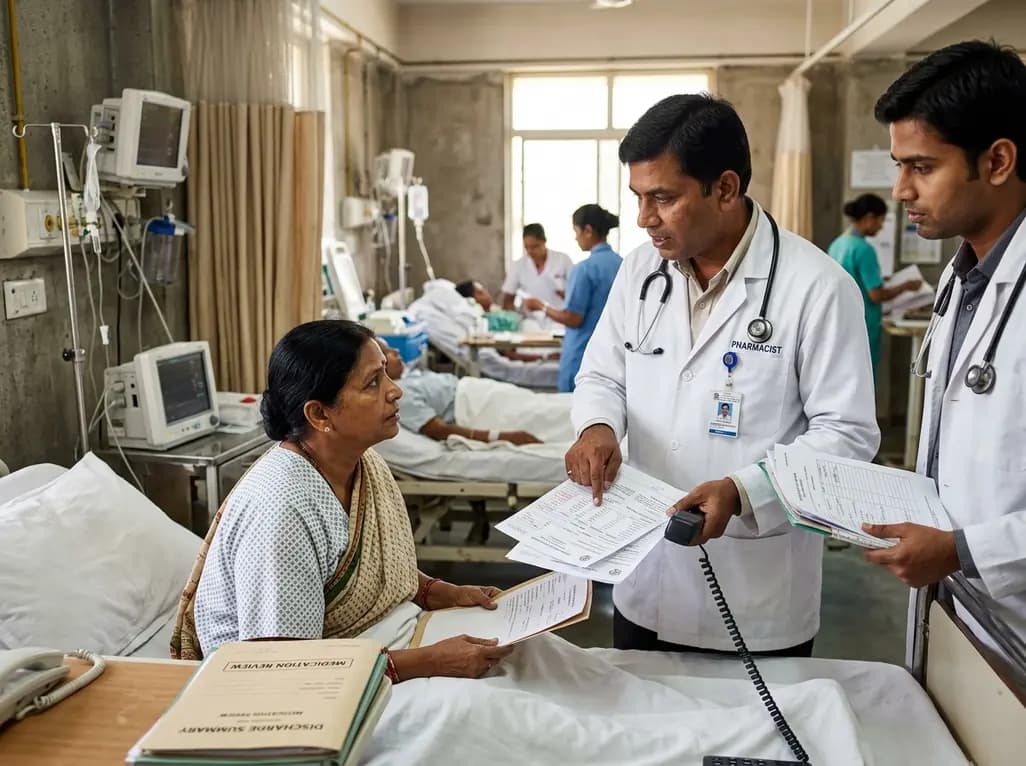
A retrospective cohort study demonstrates that comprehensive pharmacy transitions of care—including admission medication history, discharge review, and 72-hour telephone follow-up—significantly reduced 30-day readmission rates in acute care patients, highlighting the value of pharmacist-led clinical interventions.
Today

Meghalaya recorded a historic 55% reduction in maternal deaths, dropping from 244 to 107 over five years. Through the MOTHER app, dedicated transit homes, and expanded infrastructure like the 150-bed Tura Maternity Hospital with its first public NICU, the state demonstrates an effective public health model.
Today

Explore modern perioperative management protocols for clinical xenotransplantation. Learn how costimulation blockade, anti-inflammatory perfusion, and coagulopathy rescue overcome interspecies hurdles.
Today
A comprehensive bibliometric analysis evaluates 157 studies from the Swedish Spine Register (Swespine), highlighting evolving research trends, citation metrics, and the indispensable role of real-world registries in advancing spinal surgery outcomes and evidence-based practice.
Today

At Homa CME 2026 at Hyderabad's T-Hub, healthcare leaders emphasized moving beyond routine metrics through digital calculators and precision stratification. Discover why characterizing individual insulin secretion, resistance patterns, and vascular risks transforms chronic metabolic care in India.
Today
