.webp&w=384&q=75)

Loading, please wait...
Loading, please wait...

Glioblastoma multiforme represents one of the most aggressive and biologically complex primary brain malignancies encountered in neuro-oncological practice. Traditional magnetic resonance imaging primarily visualizes anatomical disruption and blood-brain barrier breakdown. Consequently, clinicians frequently face challenges when attempting to assess underlying cellular heterogeneity non-invasively. Emerging metabolic neuroimaging tools such as APTw CEST MRI now provide deeper insight into cellular protein dynamics prior to surgical resection.
Chemical exchange saturation transfer imaging exploits proton exchange between mobile solutes and bulk water. Specifically, amide proton transfer-weighted imaging detects resonance signals from amide protons within endogenous cellular proteins and peptides. Because malignant cells exhibit accelerated proliferation, they harbor significantly elevated concentrations of mobile cytosolic proteins. Therefore, the resulting CEST contrast serves as an indirect metabolic biomarker of tumor cellularity and proteomic synthesis. In contrast to conventional gadolinium-enhanced sequences, this advanced modality does not rely exclusively on microvascular leakage. Instead, it measures cellular metabolic activity directly within intact or compromised tissue architecture. Furthermore, quantitative acquisition protocols allow clinicians to map these biochemical variations across the entire intracranial mass. Consequently, neuroradiologists can differentiate viable neoplastic tissue from surrounding non-malignant vasogenic edema. Recent investigations show that this molecular approach yields valuable diagnostic information in newly diagnosed patients before therapeutic intervention. Hence, acquiring these physiological maps before surgery provides neurosurgeons and neuro-oncologists with critical baselines of biological activity. Moreover, this non-invasive assessment establishes an objective physiological foundation for subsequent longitudinal monitoring.
A recent investigation evaluated 53 adult patients with therapy-naive, IDH-wildtype glioblastoma to assess regional variations in protein saturation signals. Researchers performed preoperative scans at a median of two days prior to surgical tissue sampling. Automated deep learning-based segmentation delineated both contrast-enhancing T1 regions and surrounding FLAIR-hyperintense areas. Importantly, manual quality control validated all segmented volumes to ensure anatomic precision. The analysis revealed that APTw signal intensity was significantly higher in contrast-enhancing tumor regions than in FLAIR-hyperintense zones. This finding confirms that the core tumor mass contains substantially higher concentrations of mobile cellular peptides than the infiltrative periphery. However, the non-enhancing FLAIR margins also displayed abnormal signal profiles compared to healthy brain parenchyma. Thus, the technology successfully highlights subtle infiltrative cellular networks extending beyond the macroscopic margins seen on standard contrast images. Additionally, quantitative metrics like the 90th percentile signal intensity captured extreme biological focal points within heterogeneous neoplasms. As a result, this non-invasive profiling clarifies the true spatial distribution of proliferative glioma tissue. These observations underscore that conventional T1-weighted contrast enhancement alone fails to demonstrate the full biological extent of malignant glial invasion.
Beyond anatomical distribution, molecular subtyping dictates therapeutic responsiveness and clinical behavior in glioblastoma. The study rigorously analyzed associations between imaging parameters and established histological markers, including MGMT promoter methylation status and Ki-67 proliferative indices. Remarkably, glioblastomas assigned to the mesenchymal DNA methylation subclass demonstrated significantly higher median and 90th percentile APTw signal intensities compared to RTK1 and RTK2 subclasses. Multivariable regression models confirmed that this elevation remained statistically robust, independent of MGMT methylation status and tumor cell proliferation rates. Mesenchymal glioblastomas notoriously display aggressive invasive behavior, increased microvascular proliferation, and elevated inflammatory signatures. Therefore, the heightened signal likely reflects distinct proteomic restructuring, extracellular matrix turnover, and dense intracellular protein accumulation typical of mesenchymal phenotypes. Conversely, within the contrast-enhancing tumor compartment, elevated signal intensities showed a modest association with younger patient age. Notably, investigators found no direct, independent association between baseline APTw values and overall survival or progression-free survival. Nevertheless, these biochemical signatures successfully capture intrinsic molecular subclasses prior to surgical pathology. Consequently, non-invasive imaging profiles may help identify patients harboring the more treatment-resistant mesenchymal phenotype early in their clinical journey.
Surgical neuro-oncology relies heavily on accurate preoperative spatial mapping to maximize safe cytoreduction. Because glioblastomas exhibit profound intratumoral genetic and metabolic diversity, small tissue biopsies may inadvertently sample non-representative zones. Consequently, surgeons risk underestimating true biological aggressiveness if they sample low-grade margins. Integrating amide proton maps into preoperative neuronavigation directly mitigates this clinical sampling challenge. Specifically, neurosurgeons can fuse quantitative protein intensity maps with volumetric structural scans to identify the most biologically active tumor areas. By targeting regions displaying the highest signal percentiles, surgical teams ensure optimal tissue acquisition for essential molecular testing. Furthermore, this physiological mapping helps surgeons define margins that harbor dense cellularity despite deceptive appearances on standard T2 sequences. During resection, recognizing these active zones guides aggressive yet judicious tumor debulking near eloquent cortical structures. Therefore, advanced CEST imaging bridges the gap between gross anatomical disruption and localized microenvironmental aggressiveness. In clinical environments where neurosurgical precision directly correlates with neurological preservation, such guidance offers clear practical utility. Accordingly, surgical teams can achieve safer maximal cytoreductions while actively minimizing post-operative neurological deficits.
Beyond initial surgical staging, neuro-oncologists frequently face difficult post-treatment diagnostic dilemmas following chemoradiotherapy. Specifically, differentiating recurrent high-grade glioma from radiation-induced tissue necrosis or benign pseudoprogression remains notoriously difficult on standard imaging. Conventional contrast enhancement merely demonstrates disrupted endothelial junctions, which occurs in both recurrent malignancy and sterile treatment-related inflammation. However, metabolic imaging provides crucial clarity during this diagnostic impasse. Because recurrent active neoplasia contains rapidly dividing cells with rich cytosolic peptide pools, recurrent lesions exhibit elevated amide proton transfer values. In contrast, radiation necrosis predominantly consists of cellular debris, hypovascular fibrinoid change, and low mobile protein content, generating attenuated signals. Recent clinical trials demonstrate that combining CEST imaging with standard follow-up scans dramatically enhances diagnostic accuracy when evaluating suspicious enhancing lesions. Furthermore, this approach reduces unnecessary repeat craniotomies for pseudo-tumoral radiation changes. Clinicians can confidently tailor subsequent salvage therapy, whether requiring anti-angiogenic agents, re-irradiation, or alternative cytotoxic regimens. Thus, serial CEST evaluations offer reliable longitudinal monitoring throughout the extended continuum of glioblastoma care.
Translating advanced chemical exchange saturation transfer sequences into routine neuro-oncology workflows requires overcoming technical and institutional hurdles. Historically, extensive scan acquisition times and field inhomogeneity artifacts restricted advanced CEST sequences to dedicated academic centers. However, recent developments in 3D volume acquisition and automated B0 correction algorithms now permit reliable 3-Tesla scans within clinically feasible timeframes. Moreover, automated deep learning algorithms streamline tissue segmentation, reducing radiologist post-processing workloads while maintaining reproducible measurements. In tertiary Indian healthcare institutions and advanced neurosurgical centers, integrating these tools complements existing perfusion and diffusion modalities. Because glioblastoma management increasingly demands precision oncology, combining non-invasive metabolic profiles with next-generation sequencing data will optimize personalized therapeutic plans. Future research should evaluate whether localized protein elevations correlate with targeted therapeutic vulnerabilities, such as responses to novel immunotherapies or kinase inhibitors. As standardized acquisition packages become commercially available on standard high-field MRI platforms, clinical adoption will expand rapidly. Ultimately, non-invasive molecular characterization empowers multidisciplinary teams to deliver more refined, proactive, and individualized patient care.
APTw CEST MRI quantifies chemical exchange saturation transfer between mobile amide protons of endogenous intracellular proteins and bulk water protons. Because rapidly proliferating high-grade gliomas exhibit accelerated protein synthesis and high cellular density, they generate elevated signal intensities, allowing clinicians to assess cellular proliferation and metabolic activity non-invasively.
Recent evidence shows that glioblastomas belonging to the mesenchymal DNA methylation subclass exhibit significantly higher median and 90th percentile APTw signals than RTK1 or RTK2 subtypes. This elevated signal occurs independently of MGMT methylation status or Ki-67 labeling index, reflecting the profound proteomic alterations and aggressive microenvironment of mesenchymal tumors.
Yes, APTw CEST MRI reliably distinguishes active tumor recurrence from treatment-related necrosis or pseudoprogression. Viable, recurrent glioblastoma tissue contains densely packed cells with abundant cytosolic mobile proteins that produce elevated saturation transfer signals. Conversely, radiation necrosis consists largely of hypovascular, acellular debris, which yields noticeably lower, attenuated amide proton transfer values.
Disclaimer: This content is for informational and educational purposes only... Refer to the latest local and national guidelines for clinical practice.
References

Read summarized clinical updates, watch expert medical content, and earn CME certifications right from your smartphone.
Open your smartphone camera to scan and install.





A study reveals that APTw CEST MRI captures intratumoral heterogeneity and molecular subtypes in therapy-naive IDH-wildtype glioblastoma, showing higher signal intensities in mesenchymal tumors and contrast-enhancing regions.
Today
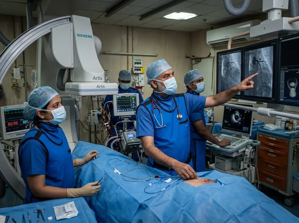
A rare case of acute coronary obstruction during TAVR caused by suspected calcified amorphous tumor embolization underscores the critical role of intracardiac imaging, emergency ECMO, and the paradox of enlarged-aperture valve geometry.
Today

Prelacteal feeding persists in South Asian communities despite known clinical risks. This review examines determinants from Chitral, pathophysiological hazards, and pediatric strategies to eliminate harmful feeds and support exclusive breastfeeding.
Today
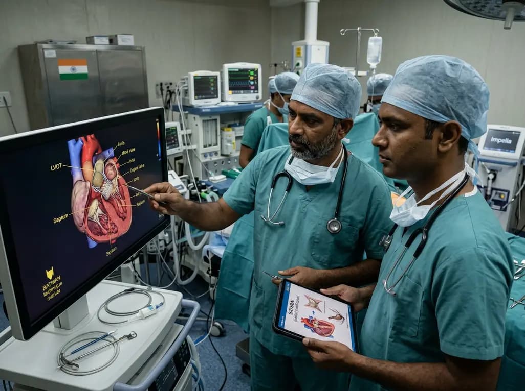
Transcatheter mitral valve replacement in patients with degenerated bioprostheses carries a substantial risk of LVOT obstruction. Combining leaflet modification via the BATMAN technique with mechanical circulatory support offers a viable, life-saving solution for anatomically challenging surgical candidates.
Today

A comparative cohort study demonstrates that patients undergoing total hip arthroplasty after prior hip arthroscopy achieve comparable functional recovery, low complication rates, and equivalent implant survivorship when conversion occurs after six months.
Today

A cross-sectional study of 320 adults evaluated the link between habitual fasting duration and dietary quality using Nutrition Quotient scores, highlighting the clinical necessity of balancing time-restricted eating with nutrient density and meal quality in routine metabolic practice.
Today
