

Loading, please wait...
Loading, please wait...

Complex craniofacial defects involving the nasal root, nasion, and anterior cranial fossa present formidable challenges for reconstructive surgical teams. Managing recurrent sinonasal inverted papilloma frequently requires radical surgical extirpation that breaches natural anatomical barriers. When conventional techniques fail, patients face persistent cerebrospinal fluid leaks, life-threatening meningitis, and extensive cosmetic deformities. In these salvage scenarios, an anterolateral thigh free flap delivers substantial vascularized tissue to re-establish intracranial-extracranial separation. Consequently, this surgical approach effectively restores structural integrity, protects vital neurological structures, and achieves predictable aesthetic outcomes.
Surgical management of recurrent sinonasal inverted papilloma demands aggressive resection because of its propensity for bone erosion and potential malignant transformation. Radical resections often leave extensive full-thickness cranial defects. These defects connect the contaminated sinonasal space directly to the intracranial cavity. Primary reconstructions traditionally rely on titanium mesh combined with vascularized local pericranial flaps. However, previous surgical manipulation, local infection, or extensive tissue loss often compromises these regional options.
When primary reconstruction fails, severe complications invariably follow. Patients frequently develop tension pneumocephalus, persistent cerebrospinal fluid leaks, and ascending intracranial infections. In addition, the presence of exposed foreign hardware exacerbates regional soft tissue necrosis and accelerates wound breakdown. Conventional local tissues cannot safely bridge such extensive voids because tension and ischemia jeopardize healing. Furthermore, the loss of nasal dorsum architecture creates noticeable contour deformities, causing profound psychological distress. Therefore, surgeons face a dual challenge: they must reliably eliminate intracranial communication while simultaneously recreating natural facial contours. Managing these refractory cases requires a disciplined reconstructive algorithm that prioritizes durable barrier formation over simple defect filling.
Free tissue transfer represents the gold standard for refractory anterior cranial defects after local reconstruction fails. Among microvascular donor options, the anterolateral thigh free flap offers unmatched versatility for cranial base salvage. First described by Song and colleagues, this reconstructive workhorse provides robust vascularity based on the descending branch of the lateral circumflex femoral artery. Moreover, its long vascular pedicle easily reaches suitable recipient vessels located outside previous surgical fields.
Surgeons can harvest the flap as a pure fasciocutaneous unit, or they can incorporate vastus lateralis muscle to obliterate irregular anatomical dead space. This adaptability proves decisive when reconstructing extensive skull base defects. The pliable fascial layer integrates seamlessly with native dural edges, promoting rapid, watertight healing. Meanwhile, the well-vascularized tissue bed actively resists residual bacterial colonization in previously contaminated fields. In addition, the donor site typically permits primary closure, which minimizes systemic donor morbidity. Consequently, the patient experiences early postoperative ambulation without major functional limitations. By supplying abundant, resilient tissue, this donor flap successfully resolves the deficiencies left by failed conventional reconstructive techniques.
A successful salvage intervention demands coordinated multidisciplinary execution by skull base neurosurgeons and microvascular reconstructive surgeons. Initially, surgeons perform a meticulous bifrontal craniotomy to re-explore the anterior skull base. The neurosurgical team removes all previously failed foreign material, debrides nonviable bone, and completely clears local inflammation. Crucially, they perform anterior sinus cranialization and mucosal exenteration to eliminate potential pathways for ascending infection.
Subsequently, the team focuses on safe pedicle routing, which represents one of the most critical surgical steps in this procedure. Traditional transfacial corridors often induce substantial facial scarring or cause pedicle compression beneath edema-prone soft tissues. Instead, surgeons route the vascular pedicle transcranially through an enlarged anterior skull base defect directly toward the temporal fossa. This clever pathway protects the delicate pedicle within a stable bony groove, shielding it from external shear forces. Next, the microvascular team performs end-to-end anastomoses to the superficial temporal vessels or facial vessels under operating microscope magnification. Because surgeons establish excellent flow with wide recipient vessels, flap perfusion remains consistently reliable. Ultimately, this thoughtful trajectory avoids skin incisions across the midface while safeguarding microvascular patency throughout the critical postoperative healing phase.
Achieving permanent intracranial-extracranial compartmentalization requires a systematic multilayered reconstructive strategy. In salvage cranial surgery, single-layer closures frequently break down under continuous pulsatile cerebrospinal fluid pressure. Therefore, surgeons utilize a comprehensive three-layer construct spanning from the dura mater to the external skin envelope. This layered approach prevents cerebrospinal fluid egress, eliminates anatomical dead space, and restores external contour simultaneously.
Specifically, the reconstructive team uses the harvested vascularized fascia lata to reconstruct the dural floor. Surgeons suture this robust fascial layer circumferentially to native dural margins using fine monofilament sutures, establishing a primary watertight barrier. Next, surgeons position vascularized muscle or subcutaneous adipose tissue between the newly formed dura and the bony framework. This bulky intermediate vascular layer obliterates cavernous dead space, cushions the frontal lobes, and prevents blood pooling. Finally, the cutaneous skin paddle resurfaces the external nasion defect and reshapes the nasal dorsum. Because the tissue maintains independent microvascular perfusion, the skin paddle heals uneventfully even in previously scarred or irradiated recipient sites. Consequently, this stratified biological seal reliably protects the intracranial contents while recreating an aesthetically pleasing nasal profile.
Clinical evidence highlights the exceptional dependability of microvascular salvage reconstruction in high-risk craniofacial defects. Staged salvage using an anterolateral thigh flap provides durable outcomes and avoids the disastrous sequelae of revision failure. Patients treated with this protocol demonstrate prompt wound healing without recurring cerebrospinal fluid rhinorrhea, pneumocephalus, or meningitis. Furthermore, the reconstruction provides stable bone support, preventing secondary enophthalmos and soft tissue contractures.
To maximize surgical success, teams must adhere to strict perioperative management protocols. Before entering the operating room, clinicians must achieve complete infection control and confirm the resolution of intracranial tension pneumocephalus. During the operation, surgeons maintain normotensive anesthesia to optimize microvascular perfusion across freshly constructed anastomoses. Postoperatively, meticulous flap monitoring through clinical checks or handheld Doppler evaluation ensures early detection of anastomotic compromise. In addition, patients undergo temporary head elevation to reduce intracranial venous pressure and minimize dependent edema. Clinicians should reserve lumbar drainage for selected cases with questionable dural integrity to prevent low-pressure headaches. Overall, combining multidisciplinary expertise with systematic technical execution transforms an otherwise catastrophic surgical failure into a stable, durable recovery.
Surgeons indicate this free flap for extensive, full-thickness anterior cranial defects when conventional local repairs fail. It is particularly valuable after infection, hardware extrusion, or pericranial flap necrosis, where robust vascularized tissue is required to prevent persistent cerebrospinal fluid leakage and seal contaminated intracranial-extracranial communications.
Surgeons route the pedicle transcranially through an enlarged skull base opening directly into the temporal fossa. This anatomical corridor shields the microvascular bundle within a bony groove, eliminating pedicle compression from facial edema. The vessels then anastomose cleanly to the superficial temporal artery and vein.
A three-layer reconstruction provides separate biological barriers spanning from deep dura to cutaneous skin. The fascial layer ensures a watertight dural seal, the vascularized muscle obliterates irregular dead space to prevent hematoma and infection, and the skin paddle reliably restores normal nasal dorsum contour and external integrity.
Disclaimer: This content is for informational and educational purposes only. It is not intended to be a substitute for professional medical advice, diagnosis, or treatment. Always seek the advice of your physician or other qualified health provider with any questions you may have regarding a medical condition. Never disregard professional medical advice or delay in seeking it because of something you have read here. Refer to the latest local and national guidelines for clinical practice.
References

Read summarized clinical updates, watch expert medical content, and earn CME certifications right from your smartphone.
Open your smartphone camera to scan and install.





Salvage reconstruction of complex anterior skull base and nasion defects after failed primary repair requires robust vascularized tissue. Staged anterolateral thigh free flap transfer with transcranial pedicle routing offers reliable intracranial-extracranial separation and restores facial contour.
Today
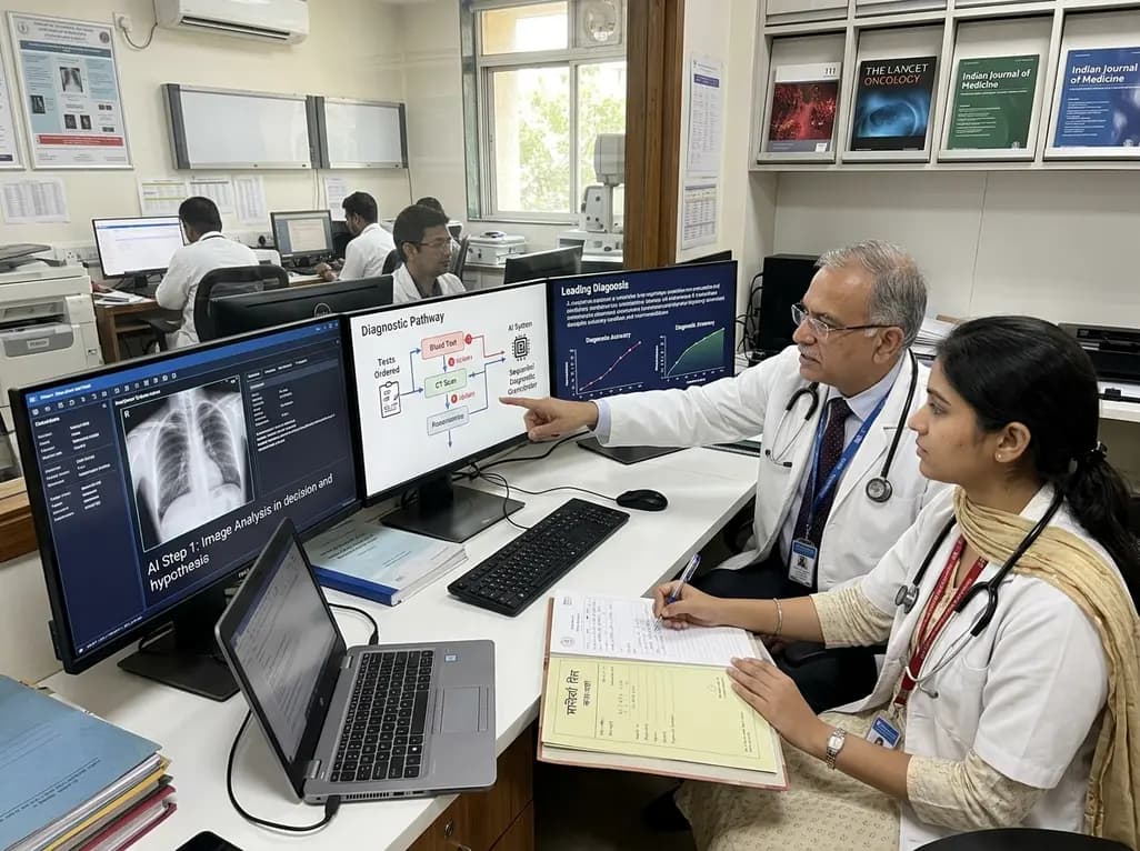
A breakthrough peer-reviewed study demonstrates that a sequential AI diagnosis system can mirror real-world clinical reasoning. By ordering stepwise tests and updating hypotheses dynamically, this multi-agent orchestrator achieved superior diagnostic accuracy while reducing investigative costs.
Today

A prenatal case highlights severe glutaric acidemia type II presenting as bilateral hyperechoic cystic kidneys at 18 weeks. Whole-exome sequencing revealed a novel homozygous ETFDH frameshift variant, demonstrating the power of rapid genetic testing in fetal renal anomalies.
Today

Review how tailored exercise programs benefit frail older patients with heart failure. Explore pathophysiological mechanisms, individualized training prescriptions, and strategies to overcome adherence barriers in clinical practice.
Yesterday

This comparative study evaluates posterior labral tears with and without glenohumeral instability, highlighting distinct demographic patterns, MRI underestimation in stable shoulders, and successful arthroscopic repair outcomes.
Yesterday

A clinical study demonstrates that early intervention with tocilizumab, either as monotherapy or bridged with corticosteroids, achieves significant proptosis reduction and resolves active inflammation in treatment-naive thyroid eye disease.
Yesterday
