.webp&w=384&q=75)

Loading, please wait...
Loading, please wait...

Developing a thorough understanding of the anterior horizontal rectus anatomy is fundamental for the successful management of pediatric strabismus. Strabismus is a condition where the eyes are misaligned, and it frequently necessitates precise surgical intervention to restore binocular vision and proper ocular alignment. Surgeons often manipulate the extraocular muscles, specifically the medial and lateral rectus, to achieve desired motor outcomes. Despite the common nature of these procedures, detailed data regarding the structural and vascular parameters of these muscles in children have historically been sparse. A recent cross-sectional study has finally shed light on the specific anatomical variations present in this demographic. By examining parameters such as anterior ciliary vessel counts, tendon width, and limbus-to-insertion distance, clinicians can gain a deeper understanding of the surgical landscape. This knowledge is particularly crucial when managing concomitant strabismus, where the deviation remains relatively constant across various gaze directions. Consequently, having a reliable baseline of anatomical norms helps in predicting surgical responses and avoiding complications such as anterior segment ischemia or unexpected overcorrections in the vulnerable pediatric population.
The vascular supply to the anterior segment of the eye is largely dependent on the anterior ciliary vessels (ACVs) that travel within the extraocular muscles. In the context of anterior horizontal rectus anatomy, the number and distribution of these vessel bundles are critical for preventing postoperative ischemia. Research indicates that ACV count patterns differ significantly between the medial rectus (MR) and the lateral rectus (LR) muscles. Specifically, the majority of lateral rectus muscles in children appear to possess two distinct ACV bundles. Interestingly, these vessel patterns do not seem to vary significantly based on the specific diagnosis of esotropia or exotropia, nor do they correlate with the magnitude of the ocular deviation. Therefore, surgeons can generally expect a consistent vascular layout when planning muscle transpositions or large recessions. Furthermore, identifying these bundles intraoperatively allows for vessel-sparing techniques, which are increasingly favored to maintain ocular perfusion. Since the vascularity remains stable regardless of the type of strabismus, the surgeon's focus can remain on the structural and positional measurements of the tendons during the procedure.
Tendon width is another essential component of the anterior horizontal rectus anatomy that influences surgical dosing and the stability of the muscle reattachment. Recent measurements in pediatric cohorts show that the mean tendon width for the medial rectus is approximately 9.98 mm, while the lateral rectus tends to be slightly narrower at 9.60 mm. These structural dimensions are not static and are influenced by various clinical variables. For instance, lateral rectus width shows a positive association with both the age of the child and the axial length of the eye. As a child grows and the globe elongates, the horizontal muscles adapt, resulting in wider tendons. This correlation suggests that surgical planning for a toddler might require different considerations compared to an adolescent. Moreover, after adjusting for age and axial length, the differences in width between different types of strabismus often become less pronounced. This finding underscores the importance of considering the overall development of the eye when evaluating muscle structure. Consequently, the surgeon must account for the physical size of the muscle tendon to ensure that the sutures are placed securely and that the force distribution across the insertion is optimal.
The limbus-to-insertion distance (LID) is a positional parameter that defines exactly where the muscle tendon attaches to the sclera. This measurement is the cornerstone of strabismus surgery, as it determines the starting point for recessions and resections. In the study of anterior horizontal rectus anatomy, the medial rectus typically displays a shorter LID, averaging 5.44 mm, compared to the lateral rectus, which averages 6.84 mm. A particularly notable finding is that the medial rectus insertion remains significantly shorter in children with esotropia even after adjusting for age and axial length. This suggests a potential anatomical predisposition or a structural adaptation related to the inward deviation of the eyes. In contrast, the lateral rectus position does not show the same level of diagnostic variation. Therefore, understanding these baseline distances is vital for accurate intraoperative measurements. If a surgeon encounters a muscle insertion that deviates significantly from these means, it may prompt a recalibration of the surgical plan to avoid under-correction or over-correction of the ocular alignment.
The integration of vascular, structural, and positional data provides a comprehensive map for pediatric surgeons. By recognizing the standard anterior horizontal rectus anatomy, clinicians can refine their surgical techniques for better predictability. For example, knowing that the medial rectus insertion is typically more anterior in esotropic patients allows for more precise recession calculations. Additionally, the awareness of ACV bundle counts aids in the safety of multi-muscle surgeries. If multiple muscles require intervention, the risk of anterior segment ischemia can be mitigated by sparing vessels in the horizontal recti. Furthermore, the association between muscle width and axial length reminds the clinician that 'one size does not fit all' in pediatric ophthalmology. Consequently, preoperative assessments that include axial length measurements may offer additional value in predicting the anatomical findings during surgery. Ultimately, this evidence-based approach to anatomy ensures that each surgical procedure is tailored to the unique physical characteristics of the child's eye, thereby improving the long-term success rates of strabismus correction.
As the pediatric eye matures, the anterior horizontal rectus anatomy undergoes significant changes that must be understood by the treating physician. The positive correlation between lateral rectus width and age suggests that the extraocular muscles grow in tandem with the orbital structures. Similarly, the axial length of the globe plays a major role in determining the dimensions and positioning of the horizontal muscles. Larger eyes with longer axial lengths tend to have wider muscle tendons, which may affect how the muscle reacts to surgical displacement. Interestingly, while the width increases with growth, the vessel patterns remain relatively constant, suggesting that the vascular architecture is established early in development. This distinction is important because it means that while the physical size of the muscle changes, the risk profile for vascular compromise may remain stable. Therefore, clinicians must remain vigilant in measuring these parameters in every case, as the interplay between growth and strabismus diagnosis creates a complex anatomical profile. By continuing to study these variables, the medical community can move toward more individualized and safer surgical interventions for children worldwide.
In children, the vascular patterns differ between the medial and lateral rectus. Research shows that approximately 59% of lateral rectus muscles possess two anterior ciliary vessel bundles. These patterns are generally consistent regardless of whether the patient has esotropia or exotropia. Understanding this distribution is essential for surgeons who wish to employ vessel-sparing techniques during strabismus surgery to maintain optimal blood flow to the anterior segment of the eye.
The limbus-to-insertion distance (LID) is a critical measure of where the muscle attaches to the eye. For the medial rectus, the insertion is typically shorter in patients with esotropia, even after adjusting for factors like age and axial length. In contrast, the lateral rectus LID tends to be more consistent across different diagnoses. This shorter medial rectus insertion in esotropia may influence the surgical strategy and the amount of recession required for correction.
Axial length is significantly associated with the physical dimensions of the extraocular muscles. Specifically, as the axial length of the eye increases, the width of the lateral rectus tendon also tends to increase. This relationship indicates that the structural features of the horizontal rectus muscles are closely tied to the overall growth and size of the globe. Surgeons should consider axial length during preoperative planning to better anticipate the muscle width they will encounter during the procedure.
Disclaimer: This content is for informational and educational purposes only... Refer to the latest local and national guidelines for clinical practice.
References
You S et al. Anterior horizontal rectus anatomy in children with concomitant strabismus. BMC Ophthalmol. 2026 Jul 13. doi: 10.1186/s12886-026-05090-3. PMID: 42443875.
Parks MM. Extraocular muscle anatomy and its clinical applications in strabismus. Ophthalmology Monographs. 2023.
Apt L. Anatomic variations in extraocular muscle insertions and the impact on surgical outcomes. Journal of Pediatric Ophthalmology & Strabismus. 2024.

Read summarized clinical updates, watch expert medical content, and earn CME certifications right from your smartphone.
Open your smartphone camera to scan and install.





A recent study characterizes the anterior horizontal rectus anatomy in children with strabismus, highlighting differences in ciliary vessel counts, tendon width, and limbus-to-insertion distance between medial and lateral rectus muscles and their clinical implications for surgical planning.
3 months ago
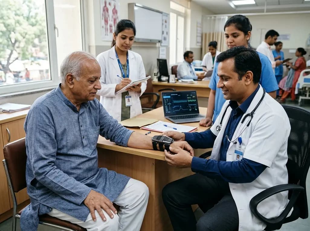
The TREM-AB open-label pilot study evaluated a wearable passive nonlinear vibration absorber for essential tremor. The Nonlinear Energy Sink bracelet reduced clinical Fahn-Tolosa-Marin tremor scores by 32.1% and electrophysiological tremor power by 49.2% with no adverse effects reported.
Today

A population-based study of 20,928 children reveals that small vulnerable newborns face sex-divergent mental capital outcomes at ages 6-7: term SGA girls show higher behavioral difficulties and lower resilience, while preterm non-SGA boys experience increased behavioral problems.
Today
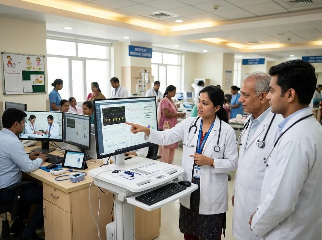
A landmark study details how an enterprise pediatric remote patient management model expanded from a single cardiology pilot to 16 diverse specialty programs by combining centralized digital operations with specialty clinical leadership.
Today
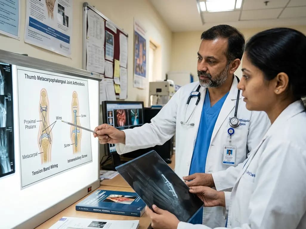
A comparative review of tension band wiring and intramedullary screw fixation in thumb metacarpophalangeal arthrodesis, evaluating union rates, hardware complications, and clinical outcomes.
Today

A breakthrough ultrathin multimodal sensor decouples physical hemodynamic monitoring from disposable electrochemical calibration, achieving long-term wearable glucose prediction with 81.36% accuracy and a 9.12% MARD. This offers a robust, drift-free solution for outpatient metabolic surveillance.
Today
