.webp&w=384&q=75)

Loading, please wait...
Loading, please wait...
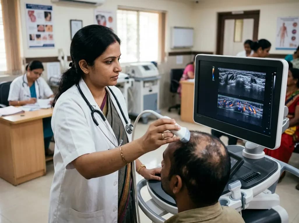
Alopecia areata presents a significant clinical challenge due to its unpredictable disease course and fluctuating inflammatory activity. Dermatologists traditionally rely on physical inspection, trichoscopy, and clinical scoring systems such as the Severity of Alopecia Tool to monitor therapeutic progress. However, these conventional modalities assess only superficial cutaneous features and cannot evaluate subclinical structural restoration or deep vascular dynamics. Recent technological advancements demonstrate that alopecia areata ultrasound imaging offers an objective, non-invasive solution to bridge this diagnostic gap. By pairing high-resolution anatomical evaluation with sensitive microvascular flow analysis, clinicians can quantify intralesional improvements well before macroscopic hair regrowth emerges.
High-frequency ultrasound operates at frequencies exceeding eighteen megahertz, which delivers exquisite spatial resolution of the upper cutaneous layers. Consequently, clinicians can clearly delineate the epidermis, dermis, and individual hair follicle structures with remarkable clarity. When clinicians integrate Superb Microvascular Imaging and power Doppler techniques, the modality overcomes the standard motion artifact limitations of conventional color Doppler. Therefore, it visualizes extremely low-velocity blood flow surrounding the follicular bulb without requiring intravenous contrast agents.
In patients with active patchy hair loss, peribulbar lymphocytic infiltrates disrupt normal microcirculation and halt the physiological hair cycle. High-frequency sonography accurately tracks these microscopic architectural alterations by recording follicular length, width, and dermal thickness. Simultaneously, spectral Doppler allows quantitative measurement of peak systolic velocity and the resistive index across cutaneous microvessels. This combined approach establishes an objective, multiparametric framework for therapeutic assessment, thereby replacing subjective visual estimation with verifiable biometric data.
Active inflammation in alopecia areata profoundly alters the structural integrity of the scalp. Sonographic analysis reveals that poorly responding or inactive follicles exhibit marked morphological degradation. Specifically, non-responding lesions display significantly shorter follicular lengths, expanded transverse diameters, and lower length-to-width ratios. These adverse dimensions directly reflect immune-mediated follicular dystropy, matrix miniaturization, and premature entry into the telogen phase.
Conversely, favorable therapeutic intervention restores follicular morphology over time. As local inflammation subsides following targeted intralesional therapy, longitudinal scans demonstrate elongation of the hair follicle shaft toward the subcutaneous tissue interface. Furthermore, dermal thickness measurements provide crucial insights into tissue remodeling and edema resolution. Ultrasound easily distinguishes healthy anagen bulbs rooted deep within the reticular dermis from miniaturized structures arrested in upper dermal levels. Thus, sequential morphological profiling serves as a sensitive surrogate marker of biological recovery.
Cutaneous hemodynamics play a pivotal role in the pathophysiology and recovery of hair follicles. Microvascular ultrasound assessment reveals distinctive hemodynamic patterns between active inflammatory states and healthy follicular regeneration. During active disease phases, intense peribulbar inflammation induces compensatory hypervascularity, which paradoxically manifests as elevated vascular index and high peak systolic velocity within affected patches.
However, successful immunosuppressive therapy resolves this pathological hyperemic state. As the peribulbar lymphocytic swarm clears, the vascular index stabilizes and localized peak systolic velocity decreases toward physiological baseline levels. Concurrently, down-regulated inflammation permits the establishment of organized, nutrient-rich capillary networks around regenerating bulbs. Therefore, tracking low-velocity perfusion parameters helps clinicians differentiate destructive inflammatory hyperperfusion from functional, restorative microcirculation during patient follow-up.
Accurate early differentiation between responders and non-responders prevents prolonged exposure to ineffective therapies and reduces cumulative drug toxicity. Patients who fail to respond to standard intralesional treatments consistently demonstrate distinct sonographic signatures. Specifically, ineffective treatment sites maintain elevated vascular indices and abnormal resistive indices alongside persistent follicular miniaturization.
In contrast, clinical responders demonstrate significant improvements across multiple sonographic parameters within weeks of initiating therapy. Their hair follicles show measurable increases in length-to-width ratios and progressive normalization of surrounding dermal echoes. Consequently, dermatologists can utilize these quantitative endpoints to refine clinical decision-making. If serial ultrasound examinations indicate persistent microvascular congestion and static follicular dimensions, clinicians can promptly modify the therapeutic regimen, switch to targeted systemic agents, or adjust dosage schedules without waiting months for visual confirmation.
Integrating high-frequency ultrasound into routine trichology practice substantially elevates the standard of patient care. The examination is rapid, repeatable, and comfortable for the patient, making it an ideal point-of-care tool. In addition, sharing objective sonographic images and quantitative vascular metrics improves patient compliance by visually demonstrating subclinical therapeutic progress.
Future developments in dermatologic ultrasound will likely incorporate three-dimensional reconstruction, automated artificial intelligence segmentation, and shear-wave elastography to assess scalp tissue stiffness. Combining these advanced modalities with standardized dermoscopic evaluation will establish a comprehensive diagnostic workflow. Ultimately, routine adoption of cutaneous sonography promises to transform alopecia management from an empirical trial-and-error process into a precise, image-guided discipline.
High-frequency ultrasound uses high acoustic frequencies to generate high-resolution cross-sectional images of the scalp. It directly measures epidermal thickness, dermal depth, and follicular length and width. Consequently, it allows clinicians to quantify structural changes, identify miniaturized follicles, and verify deep anagen regrowth before hair shafts visibly emerge through the scalp surface.
Superb Microvascular Imaging employs advanced computational algorithms to separate minute blood flow signals from tissue motion artifacts. Standard color Doppler frequently misses low-velocity microcirculation or generates excessive clutter. In contrast, this specialized technique clearly visualizes slow, fine peribulbar capillary flow, thereby enabling precise quantitative assessment of localized inflammatory and regenerative perfusion changes.
Yes, sequential ultrasound examinations provide critical early objective feedback on therapeutic response. If follow-up scans reveal persistent follicular shortening and pathological hypervascularity despite ongoing therapy, clinicians can promptly recognize treatment failure. This real-time insight allows practitioners to escalate therapy, switch medication classes, or adjust injection protocols months before clinical inspection alone confirms lack of efficacy.
Disclaimer: This content is for informational and educational purposes only and does not constitute medical advice, diagnosis, or treatment recommendations. Clinical decisions should always rely on comprehensive patient evaluation, individualized clinical judgment, and validated diagnostic protocols. Healthcare professionals should refer to the latest local and national guidelines for clinical practice.
References

Read summarized clinical updates, watch expert medical content, and earn CME certifications right from your smartphone.
Open your smartphone camera to scan and install.





High-frequency ultrasound combined with superb microvascular imaging offers an objective, non-invasive method to evaluate treatment efficacy in alopecia areata by quantifying follicular morphology and low-velocity microvascular blood flow.
last month

A laboratory technician's death in Siberia from an unverified respiratory pathogen has renewed global vigilance for pneumonic plague. Clinicians must recognize early presentations of Yersinia pestis, enforce immediate respiratory isolation, and start early bactericidal therapy to prevent severe secondary spread.
Today

Tripura has launched a comprehensive statewide oncology campaign to address late-stage breast cancer presentations. By strengthening community screening for women aged 30 and older and expanding District Day Care Cancer Centres, the initiative improves early clinical detection and decentralized chemotherapy access.
Today
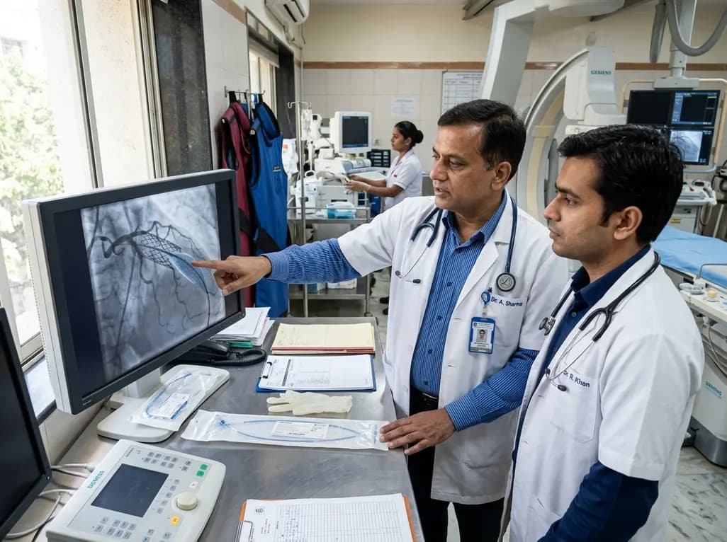
The US FDA has granted approval to the SELUTION SLR drug-eluting balloon for treating coronary in-stent restenosis. This milestone introduces a sustained-release sirolimus platform that eliminates the need for additional permanent metallic stents while maintaining robust vascular healing and luminal patency.
Today

The Thumbs-Out Classification (TOC) is a reliable screening tool for detecting thumb-in-palm deformity in children with cerebral palsy. Utilizing traffic-light tiers for radial abduction, TOC enables clinicians to monitor upper limb function and initiate timely therapeutic or surgical interventions.
Today

A comprehensive review analyzes periconceptional GLP-1 receptor agonist exposure, revealing reassuring data on congenital anomalies and adverse perinatal outcomes while emphasizing the need for cautious patient counseling.
Today
