.webp&w=384&q=75)

Loading, please wait...
Loading, please wait...

Stereotactic radiosurgery delivers ablative, highly focused radiation doses to intracranial lesions. Consequently, it represents a primary therapeutic modality for patients with secondary brain malignancies. However, clinicians frequently face a diagnostic dilemma when follow-up neuroimaging reveals an enlarging, contrast-enhancing mass with surrounding vasogenic edema. Determining radionecrosis vs tumor progression remains extraordinarily difficult because both pathophysiological entities share overlapping radiographic characteristics on conventional post-contrast T1-weighted and fluid-attenuated inversion recovery sequences. Radiation necrosis represents a delayed, immune-mediated inflammatory response accompanied by vascular fibrinoid necrosis and endothelial damage. Conversely, neoplastic progression signifies active, uncontrolled cellular proliferation demanding immediate therapeutic change. Standard clinical imaging protocols often fail to disentangle these distinct pathological processes with acceptable certainty. Therefore, neurosurgeons and radiation oncologists encounter immense uncertainty when deciding between invasive craniotomy, systemic targeted therapy, or conservative steroid regimens. An erroneous diagnosis carries significant clinical risks, including unnecessary surgical morbidity or devastating delays in oncological management.
Prior machine learning endeavors often relied on radiological follow-up or multidisciplinary consensus panels as diagnostic surrogates. However, these clinical reference standards introduce substantial verification bias and diagnostic ambiguity. In contrast, this multicenter investigation established a rigorous benchmark by analyzing 124 brain metastases with definitive histopathological validation following surgical resection. Center 1 contributed 104 lesions, whereas center 2 provided an independent cohort of 20 lesions for robust external testing. Notably, histopathological evaluation revealed pure radiation necrosis in 34 cases, representing 27.4% of the entire cohort. Neoplastic recurrence characterized the remaining lesions. By exclusively enrolling patients with definitive surgical histology, the researchers eliminated diagnostic uncertainty from the training ground truth. Consequently, this methodological rigor provided an optimal substrate for supervised computational modeling. Establishing pathological ground truth ensures that downstream neural networks and statistical classifiers learn authentic biological signatures rather than secondary imaging artifacts or ambiguous clinical impressions.
Accurate delineation of intracranial target volumes constitutes an essential prerequisite for reproducible quantitative image analysis. Manual lesion contouring remains notoriously labor-intensive and susceptible to substantial inter-observer variability. To resolve this bottleneck, investigators employed a dedicated convolutional neural network to automate the segmentation of presurgical fluid-attenuated inversion recovery and postcontrast T1-weighted sequences. The deep learning segmentation algorithm demonstrated exceptional concordance with expert manual delineations, as verified by high Dice similarity coefficients. Subsequently, the research team implemented a dual analytical architecture to evaluate diagnostic efficacy. First, they trained a Random Forest model on quantitative radiomic features extracted from 70% of the internal development cohort. Second, they engineered an end-to-end three-dimensional deep residual neural network architecture using the identical dataset split. Furthermore, the complete external dataset served strictly as an independent validation set. This dual modeling strategy permitted direct performance comparisons between feature-engineered statistical classifiers and automated end-to-end deep learning networks.
High-throughput radiomic feature extraction captured subtle phenotypic patterns invisible to the unaided human eye. Univariate and multivariate statistical analyses across the internal development cohort identified 131 significantly distinct radiomic features between necrotic and progressive lesions. Notably, textural heterogeneity metrics extracted from contrast-enhancing regions on post-contrast T1-weighted images demonstrated superior discriminative capacity. Key discriminative parameters included gray level dependence matrix dependence non-uniformity and gray level dependence matrix small dependence emphasis. These textural metrics reflect microscopic spatial variations in tissue density, microvascular breakdown, and contrast extravasation. In vital neoplastic progression, dense cellularity and irregular neoangiogenesis produce highly heterogeneous signal distribution. Conversely, radiation necrosis exhibits characteristic coagulative necrosis, vascular hyalinization, and localized liquefaction that alter parenchymal texture differently. Thus, advanced radiomic extraction effectively decodes subvisual spatial architectures, translating complex pathophysiological phenomena into reliable, quantifiable computational biomarkers for clinical evaluation.
Algorithmic performance on previously unseen external datasets represents the definitive litmus test for artificial intelligence generalizability. On the independent external test cohort, both computational architectures demonstrated exceptional diagnostic capability. Specifically, the Random Forest model attained an overall accuracy of 80.0%, an area under the receiver operating characteristic curve of 0.830, and a sensitivity of 92.8% for identifying radiation necrosis. Meanwhile, the three-dimensional deep residual neural network achieved even superior diagnostic metrics, delivering an accuracy of 85.0%, an area under the curve of 0.893, and a perfect sensitivity of 100%. The outstanding sensitivity achieved by the deep neural network holds immense clinical significance. In neuro-oncological management, false-negative evaluations for radionecrosis lead directly to avoidable surgical resections. Consequently, an algorithm that accurately rules out necrosis enables clinicians to recommend watchful observation or anti-inflammatory therapies with profound confidence, avoiding unnecessary perioperative morbidity.
Integrating artificial intelligence into routine multidisciplinary neuro-oncology workflows promises to transform post-radiosurgery surveillance significantly. Accurately distinguishing radiation effects from active neoplastic growth directly alters patient trajectories. When computational evaluation confirms true tumor recurrence, clinicians can rapidly initiate salvage therapies. These urgent interventions may include repeat focal radiosurgery, surgical decompression, or targeted systemic agents. Conversely, confirming pure radiation necrosis allows teams to implement medical treatments safely. Physicians can administer systemic corticosteroids, hyperbaric oxygen, or anti-angiogenic agents like bevacizumab without requiring diagnostic craniotomies. Additionally, non-invasive computational triage spares fragile oncology patients the functional risks, surgical stress, and logistical costs associated with exploratory surgery. As digital health infrastructures mature, deploying validated deep learning models within radiological picture archiving systems will deliver seamless, real-time diagnostic support. Ultimately, this integration elevates clinical precision, optimizes resource allocation, and substantially enhances patient quality of life across modern oncology practice.
Histological confirmation provides an absolute, objective ground truth for training algorithms. Imaging follow-up and clinical consensus often produce diagnostic errors or ambiguous classifications. Therefore, training computational models on histologically validated tissue specimens eliminates observational bias and ensures that algorithms learn authentic biological differences rather than subjective human interpretations.
Post-contrast T1-weighted and fluid-attenuated inversion recovery sequences provide the greatest discriminative utility. Enhancing areas on T1-weighted images reveal critical spatial variations in vascular integrity and tissue cellularity. Meanwhile, fluid-attenuated inversion recovery scans capture surrounding edema, which helps algorithms assess localized inflammatory responses versus infiltrative tumor growth.
Artificial intelligence models can non-invasively identify patients suffering solely from radiation necrosis with exceptional sensitivity. Consequently, neurosurgeons can safely avoid exploratory or decompressive craniotomies in asymptomatic or medically manageable individuals. This objective decision support prevents surgical morbidity, reduces healthcare expenses, and prioritizes surgery strictly for true malignant progression.
Disclaimer: This content is for informational and educational purposes only and should not be used as medical advice, diagnosis, or treatment regimens. Healthcare professionals must exercise independent clinical judgment. Refer to the latest local and national guidelines for clinical practice.
References

Read summarized clinical updates, watch expert medical content, and earn CME certifications right from your smartphone.
Open your smartphone camera to scan and install.





Differentiating radionecrosis from tumor progression after stereotactic radiosurgery is a critical clinical challenge. A multicenter study reveals that artificial intelligence and radiomics achieve high diagnostic accuracy against histopathological ground truth, potentially sparing patients invasive neurosurgery.
Today

A study evaluating drone transport of blood samples across winter and summer found 31 of 32 routine biochemical analytes remained stable compared to ground transport. While plasma LDH showed minor increases, separation gel caused greater preanalytical alterations, showing aviation rules are the main barrier.
Today

A recent case highlights favorable maternal and neonatal survival in a patient presenting at 34 weeks with probable Eisenmenger physiology and suspected double outlet right ventricle (DORV). Here is a critical review of hemodynamic challenges, delivery strategies, and peripartum management in complex cardiac disease.
Today
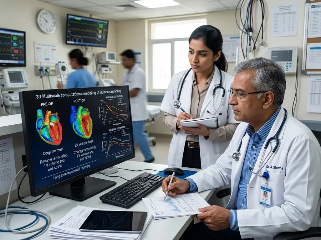
A novel multiscale computational model calibrated with Bayesian algorithms demonstrates that full reverse remodeling after mitral valve regurgitation demands both mechanical correction of volume overload and neurohormonal restoration, shedding light on persistent postoperative left ventricular dysfunction.
Today
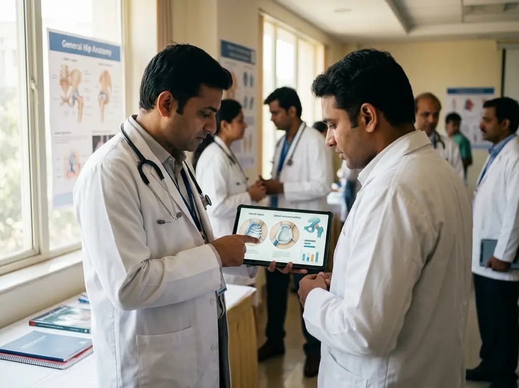
Systematic review comparing labral repair and reconstruction in primary hip arthroscopy shows comparable short- to mid-term patient-reported outcomes, with lower THA conversion after repair but superior outcomes for reconstruction in patients aged 40 and older.
Today

A preclinical study reveals that alpinetin significantly alters glimepiride pharmacokinetics by inhibiting CYP2C9. This drug-herb interaction enhances hypoglycemic potency and underscores critical safety concerns regarding severe hypoglycemia in diabetic management.
Today
