.webp&w=384&q=75)

Loading, please wait...
Loading, please wait...
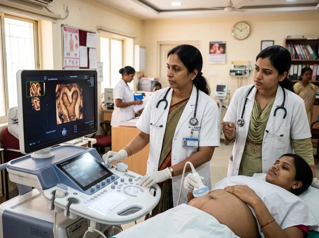
Accurate identification of congenital uterine anomalies plays a pivotal role in modern gynecological evaluation and reproductive medicine. Congenital uterine malformations arise from aberrant development, fusion, or resorption of the Müllerian ducts during embryogenesis. Consequently, affected women frequently face recurrent pregnancy loss, infertility, and preterm delivery. While three-dimensional transvaginal ultrasound represents the non-invasive gold standard, conventional assessment requires manual coronal plane reconstruction. This manual process demands considerable technical expertise and introduces operator dependency. Fortunately, emerging deep learning frameworks offer automated solutions that streamline diagnostic workflows and eliminate labor-intensive image slicing.
Congenital uterine anomalies encompass diverse structural variations, including septate, bicornuate, unicornuate, and didelphic uterine configurations. Historically, clinicians relied on hysterosalpingography and diagnostic laparoscopy to classify these anatomical deviations. However, three-dimensional ultrasound revolutionized clinical practice by visualizing both the endometrial cavity and the external uterine contour simultaneously. Expert international bodies, including the European Society of Human Reproduction and Embryology and the American Society for Reproductive Medicine, highlight the diagnostic necessity of precise coronal views. Unfortunately, acquiring an optimal coronal plane remains labor-intensive and technically challenging. Junior sonographers often struggle with acoustic shadowing, poor acoustic windows, and severe uterine retroversion. Consequently, diagnostic discrepancies frequently occur in high-volume imaging clinics. Furthermore, manual multiplanar reformatting consumes valuable clinical time during busy outpatient consultation sessions. Inexperienced operators may misclassify a partial uterine septum as a bicornuate cavity, which dangerously misguides surgical management. Therefore, reproductive specialists require reliable automated systems that diagnose congenital uterine anomalies directly from raw volumetric ultrasound data without subjective manual manipulation.
To overcome manual slicing barriers, researchers recently introduced CUA-Net, a dedicated deep learning architecture for volume classification. The investigators built the platform upon a 3-D ResNet-18 backbone specifically optimized for three-dimensional ultrasound data. Unlike traditional processing pipelines, the model processes complete volumetric inputs without requiring manual coronal plane reconstruction. Additionally, the development team integrated a dynamic data re-sampling strategy to resolve common clinical data imbalance issues. Because rare anomalies occur infrequently in clinical practice, conventional models often overfit to common anatomical presentations. CUA-Net mitigates this critical hurdle through an innovative hard sample mining technique. Specifically, this mechanism dynamically adjusts training loss to prioritize difficult and borderline anatomical geometries. Moreover, the team incorporated self-supervised volumetric reconstruction to allow the network to learn rich structural features independently. Online data augmentation further refines incorrect predictions during training iterations, thereby boosting overall diagnostic generalization. As a result, the network effectively captures intricate myometrial and cavity contours across variable clinical scanning conditions.
The research team evaluated CUA-Net across rigorous internal and external multi-center testing cohorts. In the internal validation cohort, the model delivered exceptional diagnostic metrics that confirmed its analytical stability. Specifically, CUA-Net achieved an overall accuracy of 93.88 percent and a high precision of 87.01 percent. Furthermore, the model demonstrated an impressive recall of 95.92 percent and an F-score of 88.09 percent. The micro-area under the curve reached 0.9982, while the macro-AUC achieved 0.9997. In the independent external validation set, the deep learning model maintained remarkable performance despite equipment variations. For example, CUA-Net attained an accuracy of 91.52 percent and a precision of 83.27 percent on external scans. Additionally, it sustained an 88.63 percent recall, an 81.49 percent F-score, and a macro-AUC of 0.9990. These robust findings demonstrate that the platform generalizes effectively across diverse scanner settings and patient demographics. Consequently, the algorithm resists the severe overfitting that frequently impairs earlier computer-aided ultrasound detection tools.
Clinical translation requires comparing artificial intelligence models directly against human clinicians with varying degrees of experience. In this comparative trial, CUA-Net competed against junior sonographers as well as senior ultrasound experts. Notably, CUA-Net significantly outperformed junior sonographers across every single performance indicator. Junior operators often struggle to distinguish subtle myometrial fundal notches from deep muscular septa during high-pressure clinical exams. In contrast, the intelligent model maintained precise segmentation and classification without human fatigue or perceptual bias. Moreover, CUA-Net achieved diagnostic performance that closely matched senior sonographers across most evaluated metrics. Senior specialists typically bring decades of pattern recognition experience to complex Müllerian evaluations. Matching their diagnostic precision underscores the immense practical maturity of this neural network architecture. Therefore, incorporating CUA-Net into primary triage workflows can provide junior sonographers with instant, expert-level diagnostic feedback. Such algorithmic guidance minimizes interobserver variability and elevates diagnostic confidence throughout busy obstetrics and gynecology departments.
Differentiating uterine malformations holds immense clinical significance for gynecologists, fertility specialists, and reproductive surgeons. For instance, distinguishing an arcuate or septate uterus from a bicornuate configuration directly dictates surgical intervention. Clinicians routinely treat symptomatic septate uteri using hysteroscopic metroplasty to restore normal cavity morphology. In contrast, a bicornuate uterus rarely warrants surgical reconstruction because cavity intervention does not improve live birth rates. Misdiagnosing these anomalies can expose patients to unnecessary invasive procedures or prolong heartbreaking infertility journeys. Furthermore, automated volume classification bypasses the tedious requirement of manual multiplanar manipulation during busy clinics. Clinicians can rapidly obtain reproducible structural classifications before initiating assisted reproductive technologies. Additionally, the algorithm shows promising preliminary capability in identifying rare, complex Müllerian anomalies that challenge general practitioners. Ultimately, widespread adoption of intelligent tools like CUA-Net will standardize gynecological ultrasound interpretations, reduce diagnostic delays, and substantially improve patient counseling.
CUA-Net processes complete three-dimensional ultrasound volumes directly rather than relying on manual slicing or multiplanar reconstruction. The architecture employs a 3-D ResNet-18 network that extracts volumetric spatial features across the entire organ. By utilizing dynamic data re-sampling and self-supervised volumetric learning, the model evaluates uterine morphology holistically. Consequently, it eliminates the tedious manual alignment steps that frequently introduce human error and diagnostic delays during conventional coronal plane examinations.
Differentiating a septate uterus from a bicornuate configuration directly impacts therapeutic strategy and patient safety. A septate uterus stems from incomplete resorption of the midline septum and responds favorably to hysteroscopic metroplasty, improving fertility outcomes. Conversely, a bicornuate uterus results from failed lateral fusion of the Müllerian ducts and rarely benefits from surgical repair. Misclassification can lead to inappropriate invasive surgeries or failed reproductive management, highlighting the need for accurate diagnostic tools.
Deep learning models like CUA-Net serve as decision-support systems rather than outright replacements for trained sonographers. While the model achieves diagnostic accuracy comparable to senior experts, clinicians must interpret findings within the broader clinical context. Medical history, physical examination, and concomitant pelvic pathologies remain essential for clinical decision-making. AI assistance empowers sonographers by streamlining image interpretation, reducing cognitive burden, and standardizing diagnostic quality across healthcare centers, ultimately improving patient care.
Disclaimer: This content is for informational and educational purposes only... Refer to the latest local and national guidelines for clinical practice.
References

Read summarized clinical updates, watch expert medical content, and earn CME certifications right from your smartphone.
Open your smartphone camera to scan and install.





Researchers developed CUA-Net, an intelligent deep learning framework using 3D ResNet-18 to automate the classification of congenital uterine anomalies in 3-D ultrasound without coronal plane reconstruction, achieving over 91% external accuracy and matching senior sonographers.
Today

At the New Delhi Summit, BRICS leaders adopted a landmark declaration committing member nations to united action against tuberculosis, non-communicable diseases, and future pandemic threats. Key actions include NIMHANS-led mental health networks, digital health architectures, and evidence-based integrative medicine.
Today
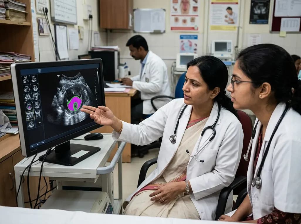
New evidence evaluates MoUNets, a static-weighted ensemble of UNet experts for automated levator hiatus segmentation in pelvic floor ultrasound. Learn how deep learning architectures perform in pelvic organ prolapse diagnostics and clinical practice.
Today

Mitochondrial deubiquitinases regulate organelle turnover by editing ubiquitin tags on damaged mitochondria. Discover how targeting these enzymes, notably USP30, offers therapeutic strategies for neurodegenerative, cardiovascular, and metabolic disorders.
Today

A rare case of a pediatric pathological femoral fracture caused by occult telangiectatic osteosarcoma demonstrates the risks of misdiagnosis after trauma, where intramedullary nailing prompted iatrogenic tumor dissemination and required modular total endoprosthetic reconstruction.
Yesterday

A cross-sectional study reveals that elevated METS-IR is independently associated with higher myopia odds in adolescents, showing biological synergy with obesity and highlighting the need for metabolic screening in pediatric eye care.
Today
