

Loading, please wait...
Loading, please wait...

Colorectal cancer remains a major global public health challenge, but screening colonoscopy significantly reduces both disease incidence and mortality. However, missed mucosal lesions continue to undermine screening efficacy in routine clinical practice. Therefore, AI assisted colonoscopy has emerged as a groundbreaking adjunct that transforms standard endoscopic mucosal visualization. Deep learning models, particularly convolutional neural networks, now analyze mucosal video streams at high frame rates. Consequently, computer-aided detection systems alert endoscopists to suspected lesions in real time. These digital visual cues draw immediate attention to subtle abnormalities that might otherwise escape human detection during withdrawal. In addition, automated algorithms help compensate for operator fatigue, perceptual variation, and visual distractions during long procedure lists. As a result, mucosal inspection becomes substantially more uniform across varied hospital environments. Endoscopy teams now view artificial intelligence not as an autonomous replacement, but as an indispensable second observer. This technological evolution marks a critical shift from experimental proof-of-concept models to validated clinical tools. Ultimately, modern gastroenterology suites increasingly utilize these intelligent systems to elevate diagnostic standards.
Substantial clinical evidence now confirms that real-time computer-aided detection consistently enhances mucosal diagnostic performance. Multiple randomized controlled trials demonstrate that automated assistance provides marked improvements in the adenoma detection rate. In addition, these multicenter investigations report significant increases in overall polyp detection rates and adenomas identified per colonoscopy. Most notably, artificial intelligence effectively lowers the adenoma miss rate by highlighting overlooked lesions along mucosal folds. Recent clinical data indicate that algorithmic benefits appear especially pronounced for diminutive polyps measuring five millimeters or less. Furthermore, the technology successfully flags subtle flat and sessile serrated lesions that human eyes easily bypass. However, the exact clinical benefit varies according to operator experience and baseline diagnostic skill. Less experienced endoscopists often experience greater performance gains compared to seasoned high-detecting practitioners. Similarly, local bowel preparation quality substantially influences algorithm accuracy and sensitivity. Clean mucosal surfaces allow detection models to function with superior precision during systematic withdrawal. Therefore, gastroenterologists must maintain meticulous cleaning techniques while leveraging digital assistance. By integrating these systems into daily practice, endoscopy units establish a reliable safeguard against diagnostic oversights.
Although artificial intelligence clearly improves surrogate detection benchmarks, fundamental epidemiological questions remain unanswered. Traditionally, an elevated adenoma detection rate strongly correlates with a lower incidence of interval colorectal cancer. Nevertheless, researchers still debate whether incremental gains driven primarily by diminutive polyps confer true long-term oncological protection. Small adenomas exhibit exceedingly low rates of malignant progression over standard screening intervals. Consequently, identifying dozens of tiny benign lesions may simply inflate administrative quality scores without decreasing cancer mortality. In addition, the increased detection of low-risk polyps generates substantial procedural consequences for healthcare organizations. Endoscopists must perform more polypectomies, which slightly increases the risks of post-procedural bleeding and colonic perforation. Furthermore, finding more lesions forces patients into tighter colonoscopic surveillance intervals under prevailing post-polypectomy guidelines. This dynamic increases direct healthcare expenditures and strains already overburdened endoscopy capacity. Therefore, major medical societies recommend cautious, thoughtful adoption rather than uncritical clinical reliance. Future longitudinal trials must confirm whether computer-assisted mucosal detection genuinely suppresses post-colonoscopy colorectal cancer rates. Until such robust survival data emerge, clinicians should balance enthusiasm with rigorous clinical judgment.
While detection tools command significant attention, researchers have concurrently developed computer-aided diagnosis to characterize mucosal lesions. Modern CADx architectures analyze surface microvasculature and pit patterns using advanced optical chromoendoscopy and white-light modalities. By evaluating these microscopic morphological features, diagnostic algorithms differentiate neoplastic adenomas from nonneoplastic hyperplastic polyps in real time. Consequently, this capability supports paradigm-shifting management approaches, including the resect-and-discard strategy for diminutive rectosigmoid lesions. Furthermore, endoscopists could potentially leave benign hyperplastic polyps in place without tissue resection, known as diagnose-and-leave. Adopting these optical biopsy pathways could drastically reduce pathology processing costs and avoid unnecessary intervention risks. However, clinical implementation of characterization algorithms remains far less mature than computer-aided detection systems. Most characterization models require high-confidence diagnostic thresholds and pristine image quality to ensure accurate optical classification. Misclassifying an advanced neoplastic lesion as benign carries grave oncological risks for patients undergoing routine surveillance. In addition, current medicolegal frameworks often mandate formal histological confirmation before altering surveillance recommendations. As a result, gastroenterologists currently utilize diagnostic assistance primarily as educational adjuncts while awaiting broader regulatory clearance.
Successfully embedding artificial intelligence into busy clinical workflows requires addressing several practical challenges. Frequent false-positive notifications represent a major operational hurdle during mucosal examination. Normal mucosal folds, retained stool fragments, and suction artifacts can trigger erroneous visual bounding boxes. Consequently, frequent alerts can induce alert fatigue, which distracts practitioners and subtly prolongs withdrawal times. Moreover, clinicians must guard against operator deskilling and dangerous overreliance on digital tools. Endoscopists who depend excessively on automated prompts might compromise their independent mucosal inspection technique. Therefore, training programs must emphasize that software operates strictly as a supportive aid rather than a replacement for meticulous technique. In addition, healthcare facilities face notable hardware procurement costs and interoperability hurdles across proprietary endoscopy platforms. Seamless integration demands user-friendly interfaces that preserve ergonomic comfort and procedure efficiency. Fortunately, modern systems offer flexible display settings, allowing operators to adjust alert sensitivity according to clinical preference. Ultimately, artificial intelligence will achieve its full potential only through human-centered design. When endoscopists synthesize automated insights with clinical judgment, overall procedural quality reaches new heights.
Computer-aided detection focuses primarily on locating polyps on the colonic mucosa by displaying real-time visual bounding boxes around suspicious tissue. In contrast, computer-aided diagnosis characterizes detected lesions by analyzing surface vascular patterns and pit architecture. This diagnostic differentiation categorizes polyps as neoplastic adenomas or benign hyperplastic tissue. Consequently, computer-aided detection increases lesion identification, whereas computer-aided diagnosis guides clinical decisions regarding endoscopic resection and pathology submission.
While higher baseline adenoma detection correlates with lower interval cancer rates, direct survival evidence remains unconfirmed for automated systems. Clinical trials show that software predominantly detects diminutive polyps measuring under five millimeters. However, these tiny lesions carry minimal short-term malignant risk. Therefore, researchers need long-term studies to prove that algorithmic gains genuinely reduce colorectal cancer incidence and mortality over time. Furthermore, health economic registries will evaluate surveillance burdens.
Endoscopists primarily confront false-positive alerts that disrupt withdrawal flow and cause cognitive alert fatigue during procedures. In addition, acquiring specialized processing hardware incurs substantial capital costs for endoscopy units. Clinicians also face the risk of operator deskilling if they rely uncritically on automated bounding boxes. Therefore, professional guidelines recommend using artificial intelligence strictly as an assistive second reader while maintaining rigorous, independent mucosal inspection techniques. Furthermore, robust training curricula ensure standard diagnostic proficiency across teams.
Disclaimer: This content is for informational and educational purposes only... Refer to the latest local and national guidelines for clinical practice.
References

Read summarized clinical updates, watch expert medical content, and earn CME certifications right from your smartphone.
Open your smartphone camera to scan and install.





Artificial intelligence is transforming colonoscopy through computer-aided detection (CADe) and diagnosis (CADx). While AI significantly boosts adenoma detection rates, ongoing research is evaluating its direct impact on long-term colorectal cancer mortality and cost-effective clinical implementation.
Today
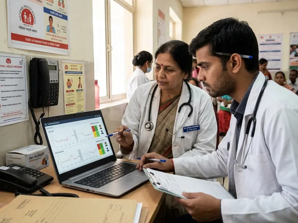
A novel adherence-driven sparsity framework demonstrates that missingness in patient-generated telemedicine data reflects human behavior rather than mere technical error, guiding safer clinical interpretations.
Today
Arthrogryposis multiplex congenita affects fetal joint mobility, yet fewer than half of cases are detected prenatally. This review highlights dynamic fetal motor assessment, phenotypic subtyping, rapid exome sequencing, and multidisciplinary perinatal care to optimize diagnosis and parental counseling.
Today
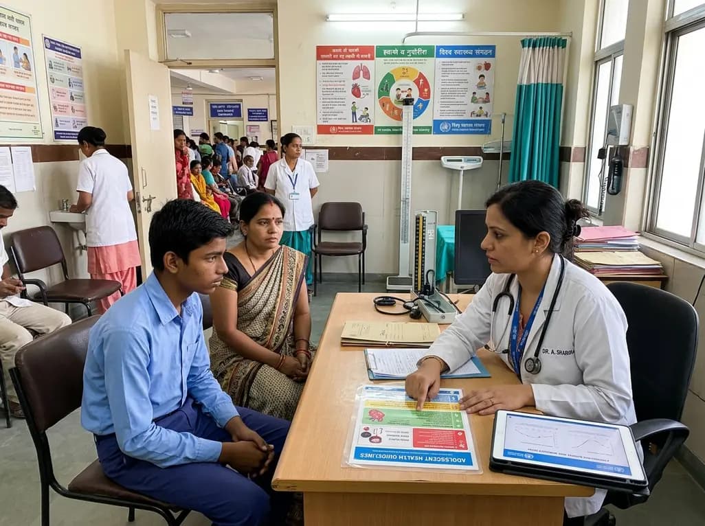
A major World Health Organization study reveals that one in four teenagers globally records zero active days per week. Discover the epidemiological data, cardiometabolic consequences of adolescent sedentary behavior, and practical guidance for clinicians to promote healthy lifestyle habits among youth.
Today
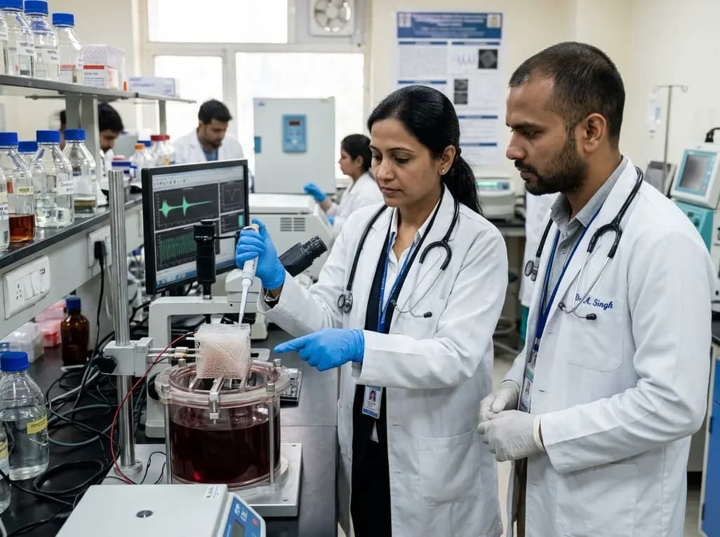
Engineered 3D piezoelectric PHBV scaffolds generate electrical charges during mechanical strain, mimicking native muscle electromechanics to promote cellular alignment, myogenic differentiation, and functional tissue repair in severe volumetric muscle loss.
Today
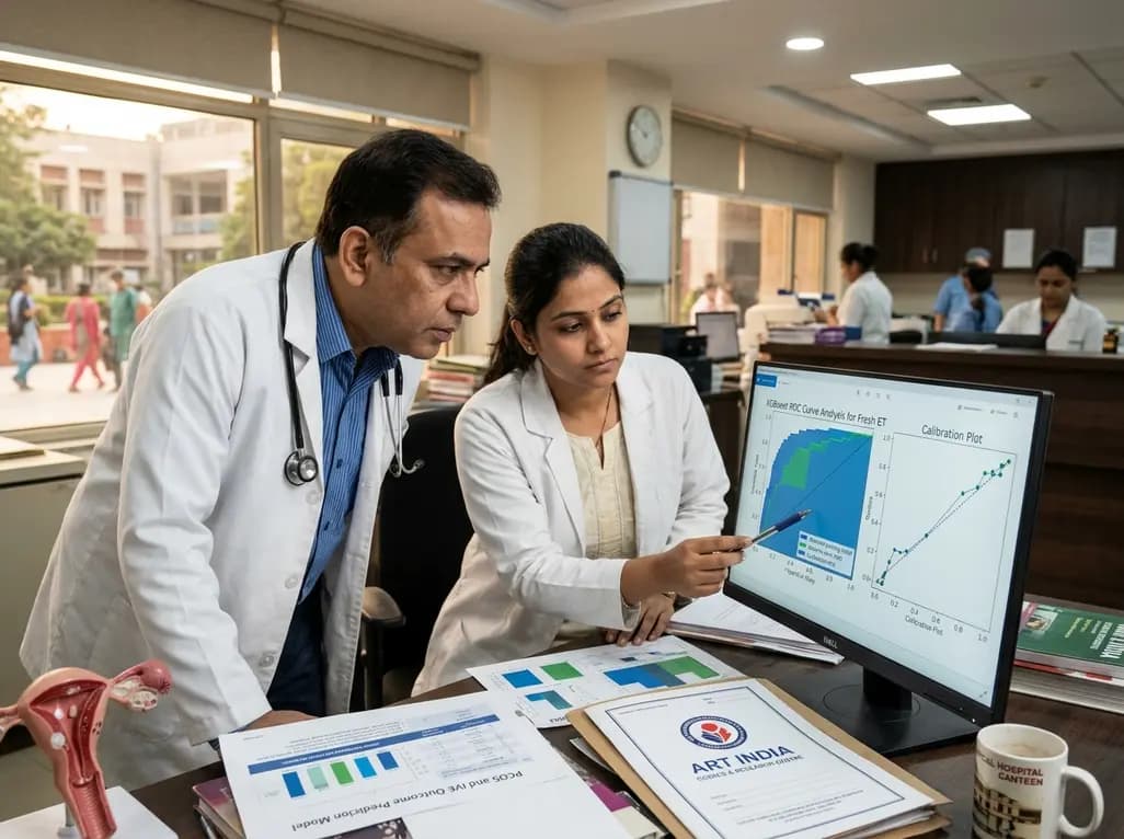
A multicenter study evaluates an XGBoost machine learning model to predict miscarriage risk in PCOS after fresh embryo transfer. While discrimination was strong internally, cross-cohort calibration drift emphasizes its role as a risk-ranking tool rather than a standalone diagnostic instrument.
Today
