

Loading, please wait...
Loading, please wait...
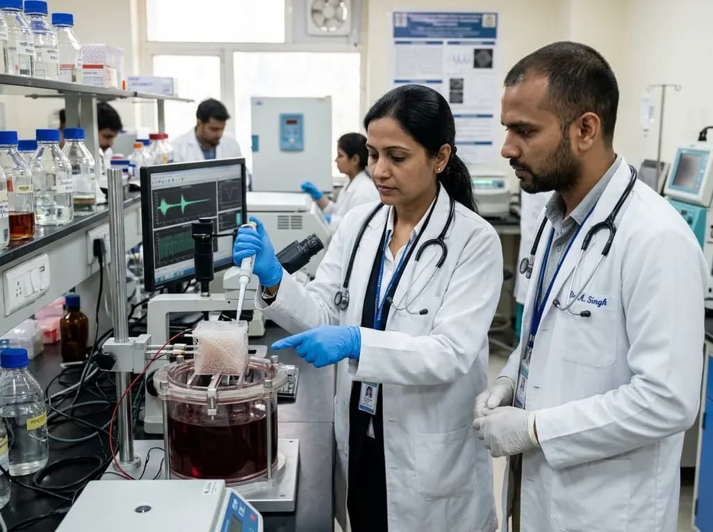
Severe skeletal muscle trauma and volumetric muscle loss present formidable clinical challenges for orthopedic and reconstructive surgeons. Standard autologous muscle flaps often cause donor-site morbidity, while passive synthetic implants fail to restore contractile function. Fortunately, recent breakthroughs in bioresorbable piezoelectric scaffolds offer transformative therapeutic opportunities. These biomimetic fibrous matrices generate intrinsic electric charges during movement, emulating the native electromechanical environment of contracting muscle. Consequently, they provide essential biophysical cues that promote cell migration and functional tissue regeneration.
Skeletal muscle possesses an inherent capacity for spontaneous repair after mild injury. However, severe high-energy trauma, tumor ablations, and blast injuries often produce massive volumetric muscle loss. In these catastrophic circumstances, endogenous regeneration fails completely because structural architecture is missing. Fibrotic scar tissue rapidly infiltrates the defect, which severely impairs mechanical function and produces permanent disability. Currently, surgeons rely on autologous free muscle transfers to manage extensive defects. Although free tissue transfer provides vascularized soft tissue coverage, it inevitably creates significant donor-site weakness. Furthermore, standard passive biomaterials provide only structural support without delivering active regenerative cues. Native skeletal muscle is an electroactive tissue that depends on coordinated electromechanical signals during development and repair. Therefore, passive acellular scaffolds cannot stimulate functional myofiber alignment or restore muscle strength. Clinicians require advanced regenerative matrices that supply both structural frameworks and physiological electrical stimulation. By addressing these critical limitations, biomimetic tissue engineering introduces powerful strategies to rebuild damaged skeletal muscle.
To bridge the therapeutic gap, researchers engineered biomimetic piezoelectric scaffolds using poly(3-hydroxybutyrate-co-3-hydroxyvalerate), known as PHBV. This natural biopolymer exhibits excellent biocompatibility, predictable biodegradability, and inherent piezoelectric properties. When mechanical stress deforms the scaffold, internal molecular dipoles generate electrical surface charges without requiring batteries or wires. Consequently, everyday patient movement and cyclic physical therapy produce dynamic electrical microcurrents directly within the wound bed. This electromechanical activity accurately mimics the endogenous bioelectric environment of contracting muscle bundles. In addition, PHBV undergoes progressive enzymatic hydrolysis, releasing harmless metabolites like hydroxybutyrate that nearby cells easily metabolize. Therefore, the polymer avoids chronic foreign-body inflammation commonly observed with non-degradable piezoelectric materials like polyvinylidene fluoride. Furthermore, dynamic surface electrical charges activate cell membrane channels, which dramatically stimulates cellular adhesion and proliferation. Ultimately, PHBV matrices combine structural elasticity with self-powered electrical cues to guide functional muscle repair.
Fabricating successful tissue scaffolds requires precise optimization of structural and electrical characteristics. In recent studies, bioengineers used advanced electrospinning technology to produce three-dimensional fibrous PHBV constructs. Electrospinning produces nonwoven microfibrous architectures that closely resemble natural extracellular matrix morphology. Specifically, investigators evaluated how solvent mixtures, polymer concentrations, and tip-to-collector distances influence fiber diameter and porosity. By systematically optimizing these fabrication parameters, the researchers achieved controlled pore connectivity and superior electromechanical responses. Notably, optimized scaffolds showed a twenty-five-fold surge in the quasi-static piezoelectric coefficient, rising from 0.117 to 2.9 pC/N. Dynamic Vector Network Analyzer measurements confirmed an electromechanical coupling factor of 13.86%, indicating robust energy conversion efficiency. Furthermore, mechanical testing proved that these fibrous constructs withstand tensile strains up to 40% before failure. Because native skeletal muscle contracts dynamically, scaffold compliance remains vital to prevent structural failure. Thus, electrospun PHBV constructs provide an ideal balance of mechanical flexibility and electroactive responsiveness.
The ultimate value of any tissue construct depends on its biological compatibility and cellular influence. In cell culture experiments, electrospun PHBV scaffolds supported robust adhesion and proliferation of myogenic progenitor cells. The interconnected microporous network facilitated rapid nutrient transport and metabolic exchange across the fibrous mesh. More importantly, preliminary assays revealed that piezoelectric stimulation actively promotes myogenic differentiation. When muscle progenitor cells detect local surface charges, their voltage-gated ion channels open immediately. Consequently, intracellular calcium influx increases, which stimulates essential myogenic regulatory genes including MyoD and myogenin. In addition, aligned fibrous topographies guide elongated myoblasts to align in parallel arrays and fuse into multinucleated myotubes. In contrast, passive culture substrates lack electrodynamic signaling, often resulting in disorganized cell growth. Therefore, the electromechanical coupling of PHBV converts mechanical motion directly into biological differentiation signals. Ultimately, this coordinated electromechanical stimulation accelerates myogenesis, creating functional muscle bundles suitable for active contraction.
These pioneering biomaterial advancements hold immense translational promise for trauma care, orthopedics, and sports medicine. Severe road traffic accidents, occupational crush injuries, and complex sports trauma frequently produce challenging soft-tissue defects. By implanting resilient piezoelectric scaffolds, reconstructive surgeons can bridge substantial muscle gaps without harvesting healthy autologous muscle flaps. Because PHBV constructs accommodate strains up to 40%, they easily flex alongside host tissues during movement. Furthermore, postoperative physical rehabilitation directly triggers piezoelectric surface charges, transforming physical therapy into an active biochemical regenerative driver. In addition, the degradation profile of PHBV synchronizes with cellular ingrowth, preventing sudden mechanical failure during tissue maturation. Surgeons could also combine these smart scaffolds with autologous progenitor cells or growth factor coatings to accelerate functional integration. However, investigators must conduct rigorous large-animal trials to verify long-term vascular perfusion and functional motor innervation. Ultimately, self-powered piezoelectric biomaterials represent a paradigm shift in musculoskeletal reconstructive surgery, paving the way toward scarless functional restoration.
Piezoelectric scaffolds utilize the natural molecular properties of polymers like PHBV to convert mechanical deformation into localized electrical charges. Consequently, standard physiological movement or physical rehabilitation flexes the scaffold fibers, which instantly produces transient surface voltages. Furthermore, this dynamic electromechanical response mimics native muscle depolarization. As a result, nearby myoblasts receive continuous biophysical stimulation, accelerating cellular alignment and myotube fusion without requiring transcutaneous wiring or implanted batteries.
Poly(3-hydroxybutyrate-co-3-hydroxyvalerate), or PHBV, combines biocompatibility and predictable bioresorption with inherent piezoelectricity. Unlike inert materials that merely provide structural frameworks, PHBV generates active electrical dipoles under mechanical strain. In addition, its degradation produces non-toxic metabolites that tissues easily process without excessive inflammation. Therefore, PHBV scaffolds provide mechanical structural support while actively delivering regenerative electromechanical cues, significantly outperforming passive synthetic polymers in promoting functional muscle tissue regeneration.
Although laboratory investigations demonstrate excellent cytocompatibility, several surgical translational challenges remain unresolved. Specifically, researchers must evaluate long-term vascularization and functional nerve innervation in large-animal defect models. Furthermore, surgeons require standardized surgical fixation techniques for irregular wound beds. Clinical teams must also validate degradation kinetics against human tissue healing rates. Consequently, establishing standardized Good Manufacturing Practice production and thorough regulatory verification remain critical before human clinical trials can begin.
Disclaimer: This content is for informational and educational purposes only and does not constitute medical advice, diagnosis, or treatment. Healthcare professionals should exercise independent clinical judgment. Refer to the latest local and national guidelines for clinical practice.
References

Read summarized clinical updates, watch expert medical content, and earn CME certifications right from your smartphone.
Open your smartphone camera to scan and install.





Engineered 3D piezoelectric PHBV scaffolds generate electrical charges during mechanical strain, mimicking native muscle electromechanics to promote cellular alignment, myogenic differentiation, and functional tissue repair in severe volumetric muscle loss.
Today

A recent study highlights that pairing photograph-assisted 24-hour recalls with regional food databases significantly boosts dietary assessment accuracy. This methodology reduces cultural bias and strengthens nutrient tracking, offering crucial lessons for clinicians managing nutritional care in diverse populations.
Today

A preclinical study reveals that cinnamic acid counters D-galactose-induced premature ovarian failure in mice. By enhancing total antioxidant capacity and upregulating protective genes like Nrf2 and Foxo3a, cinnamic acid improved estrous cyclicity and follicular reserve, offering new insights into ovarian health.
Today

Wearable e-textiles transform cardiovascular monitoring by integrating flexible conductive sensors into everyday clothing. This advance enables continuous, non-invasive assessment of ECG, pulse waves, and hemodynamics, bridging the gap between hospital diagnostics and home healthcare.
Today

India's food safety authority is taking aggressive action against major beverage companies for marketing high-caffeine products as energy drinks. This clinical analysis examines the cardiovascular complications, metabolic risks, and pediatric concerns behind the regulator's push for truthful beverage standards.
Today
