

Loading, please wait...
Loading, please wait...

Cognitive decline presents an escalating clinical challenge worldwide, particularly across super-aged societies and resource-constrained healthcare systems. Traditional neurocognitive screening relies heavily on active pen-and-paper assessments such as the Mini-Mental State Examination. However, these conventional batteries frequently suffer from cultural biases, education-dependent disparities, and test fatigue. Consequently, investigators are exploring digital phenotyping as a scalable, noninvasive diagnostic alternative. Recent research indicates that quantifying spontaneous affective facial responses through artificial intelligence provides an objective behavioral marker for staging neurodegenerative conditions. By evaluating how older adults involuntarily react to emotional stimuli, clinicians can glean critical insights into early frontotemporal and subcortical functional decline.
Emotional processing involves intricate interactions between frontal cortical circuits, the limbic system, and brainstem cranial motor nuclei. Spontaneous affective facial responses reflect real-time emotional reactivity and sensory integration. When neurodegenerative pathology compromises neural tracts, expressivity and affective processing inevitably deteriorate. In conditions like mild cognitive impairment and overt dementia, pathological deposition of amyloid and tau proteins disrupts the amygdalohippocampal complex and frontal circuitry. Consequently, affected individuals frequently manifest emotional blunting, delayed facial expression initiation, or incongruent responses to emotional triggers. Neuroscientists recognize that involuntary emotional mimicry occurs automatically via mirror neuron networks. Therefore, alterations in facial dynamics can reveal micro-level neurobehavioral changes long before gross functional disability emerges. Clinicians can harness these micro-expressions as reliable behavioral windows into neurological integrity.
To quantify dynamic facial behavior objectively, researchers developed computer vision pipelines utilizing convolutional neural networks. These deep learning systems evaluate two core digital metrics: affective expression congruency and onset latency. Congruency denotes whether the participant's facial expression matches the valence of the presented emotional video. For instance, displaying smiling geometry during an amusing clip denotes congruency, whereas a blank mask or a grimace represents neutrality or incongruence. On the other hand, onset latency calculates the exact temporal duration from stimulus onset to the emergence of detectable emotional facial movement. Automated facial action coding systems capture these fleeting muscular activations with high frame-rate precision. Consequently, algorithmic monitoring eliminates subjective examiner bias and delivers granular, reproducible behavioral metrics across diverse clinical settings.
A landmark exploratory feasibility study revealed striking disparities in facial reactivity among healthy older adults, patients with mild cognitive impairment, and individuals with dementia. During brief positive stimuli, healthy controls exhibited congruent positive facial expressions in 65% of cases. Conversely, individuals with mild cognitive impairment displayed congruent expressions in only 20% of trials, while patients with dementia demonstrated an absolute absence of congruent responses. Moreover, negative emotional stimuli elicited congruent negative expressions in 60% of healthy participants, compared to a mere 5% among both cognitive impairment cohorts. Furthermore, affective onset latency showed marked elongation in cognitively impaired cohorts. Healthy adults responded swiftly, whereas individuals with advancing pathology demonstrated prolonged delays or failed to mount any visible response. Even after statistical adjustment for age and formal education, these diagnostic divergences remained highly significant.
Integrating video-based facial analytics into existing outpatient workflows offers distinct logistical advantages. First, the noninvasive capture protocol requires only a standard digital camera or consumer-grade tablet device. Patients passively observe brief video vignettes while completing routine cognitive tasks. Therefore, the assessment minimizes clinical anxiety and bypasses educational barriers that commonly skew paper-based psychometric testing. Second, automated scoring provides rapid triage data, allowing geriatricians and neurologists to flag subtle neurobehavioral deficits during primary care encounters. In community settings and memory clinics, such digital biomarkers can identify vulnerable individuals who warrant comprehensive diagnostic workups. Clinicians can subsequently prioritize high-risk patients for targeted neuroimaging, cerebrospinal fluid analysis, or emerging plasma biomarker panels.
Although computer vision offers promising screening utility, physicians must interpret current findings within an exploratory framework. Facial expressivity naturally varies across different ethnic backgrounds, baseline personality profiles, and concurrent neuropsychiatric states. For example, late-life depression and parkinsonian masking can mimic blunted emotional reactivity. Furthermore, neuromuscular disorders or peripheral nerve palsies may introduce diagnostic confounding. Machine learning algorithms also require rigorous multi-center validation across ethnically diverse cohorts to prevent algorithmic bias. Technical considerations such as ambient room illumination, participant head orientation, and visual acuity must also be standardized. Nevertheless, combining automated facial analysis with speech analytics and digital motor tracking holds tremendous promise for comprehensive multimodal digital phenotyping in neurodegenerative disease management.
Affective facial responses represent spontaneous micro-expressions generated by patients when viewing emotion-provoking stimuli. AI algorithms track muscle activations to evaluate emotional congruency and reaction latency. In dementia evaluations, these metrics provide an objective digital measure of underlying affective processing. They reveal subtle impairments within frontolimbic circuits that govern emotional comprehension and spontaneous motor expression.
Artificial intelligence uses deep convolutional neural networks trained on extensive facial datasets to identify specific action units. These systems map critical landmarks around the eyes, eyebrows, and mouth at millisecond intervals. Consequently, the software accurately detects subtle shifts in affective valence, measuring whether the response matches the stimulus and timing how quickly the movement begins.
Facial analysis cannot replace comprehensive neuropsychological assessments or biomarker testing. Instead, clinicians utilize these digital tools as noninvasive, scalable screening adjuncts. They assist in identifying individuals who exhibit early neurobehavioral alterations, enabling timely referral for confirmatory evaluations, including detailed psychometric batteries, advanced neuroimaging, and molecular biomarker testing to establish a definitive diagnosis.
Disclaimer: This content is for informational and educational purposes only and should not be considered medical advice. Always consult a qualified healthcare provider for diagnosis and treatment decisions. Refer to the latest local and national guidelines for clinical practice.
References
Yang JC et al. AI-Assisted Analysis of Spontaneous Affective Facial Responses for Cognitive Status Assessment in Older Adults: Exploratory Feasibility Study. JMIR Form Res. 2026 Nov 02. doi: 10.2196/94422. PMID: 42970778.
Al-Nuaimi AH et al. AI-Based Facial Emotion Analysis for Early and Differential Diagnosis of Dementia. Bioengineering (Basel). 2025 Oct 04;12(10):1082. doi: 10.3390/bioengineering12101082.
Ferrer-Cairols I et al. From cognitive screening to digital phenotyping: rethinking early detection of cognitive impairment in primary care. Front Psychiatry. 2025;16:1428901. doi: 10.3389/fpsyt.2025.1428901.

Read summarized clinical updates, watch expert medical content, and earn CME certifications right from your smartphone.
Open your smartphone camera to scan and install.





A novel exploratory study highlights the feasibility of using AI-derived metrics of spontaneous affective facial responses to differentiate healthy older adults from those with mild cognitive impairment and dementia, offering an objective, scalable digital biomarker for early neurocognitive assessment.
Today
Standardizing continuous physiological waveforms in EHRs has long challenged clinical teams. The novel TIDE framework leverages HL7 FHIR R4 to decouple queryable clinical metadata from massive raw signal repositories, unlocking scalable telemetry exchange for bedside care and predictive research.
Today

A secondary analysis of the MOMS clinical trial reveals that infant sex does not influence neonatal opioid withdrawal syndrome severity in buprenorphine-exposed newborns. Maternal co-exposures and clinical factors drive outcomes, highlighting the need for objective assessment and supportive breastfeeding.
Today
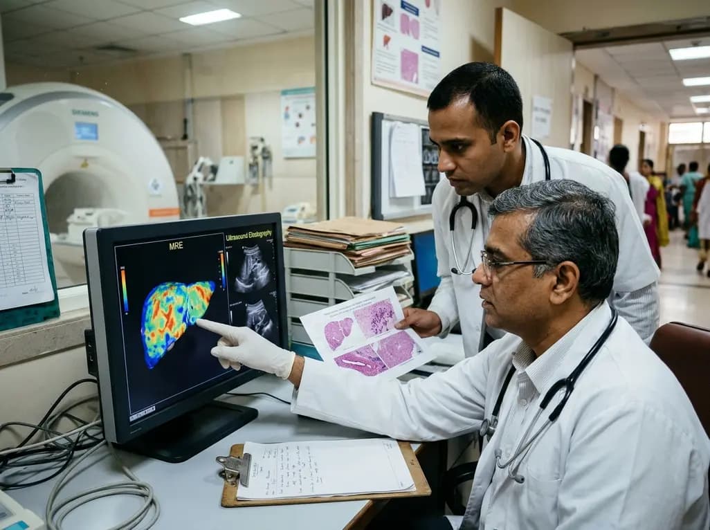
A comparative study reveals magnetic resonance elastography outperforms ultrasound elastography in assessing liver fibrosis in Fontan-associated liver disease, demonstrating stronger histological agreement and superior discrimination.
Today
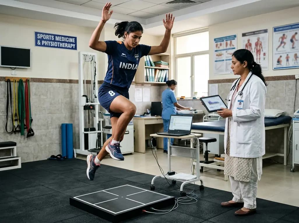
A systematic review and meta-analysis highlights the substantial impact of plyometric jump training on jump performance, reactive strength, and landing kinematics in female team-sport athletes, providing evidence-based programming guidelines for sports medicine and orthopedics.
Today

A comprehensive review of the role of growth hormone in diabetic kidney disease and podocytopathy. Learn how elevated GH activates ZEB2, Notch, and TGF-beta1, driving podocyte loss and glomerulosclerosis, and explore novel targeted therapeutic approaches.
Today
