.webp&w=384&q=75)

Loading, please wait...
Loading, please wait...

Intracranial pial AVFs are rare cerebrovascular shunts. In these lesions, pial arteries connect directly to veins without an intervening nidus. Because these lesions are uncommon, clinicians often find their anatomical features difficult to understand. However, a recent multicenter study by Yamada et al. proposes a new intracranial pial AVF classification based on angioarchitecture to improve clinical diagnosis.
Researchers conducted a nationwide retrospective cohort study in Japan between 2013 and 2024. They centrally reviewed imaging data to confirm diagnoses and categorize lesions. Specifically, the team focused on adult cases to better understand the clinical implications of these rare shunts. Consequently, the study provides comprehensive data on adult pial AVFs and their unique vascular structures.
One major advancement in this study is the definition of microfistulas. The authors identified these lesions by the small diameter of the shunt feeder. Furthermore, the angioarchitectural classification helps pinpoint specific feeder patterns that dictate treatment choice. For instance, distinguishing between single-feeder and multiple-feeder systems is crucial for procedural success. Additionally, the classification system accounts for venous drainage patterns and the presence of venous pouches.
Treatment for pial AVFs usually involves endovascular embolization or microsurgical ligation. Because these fistulas have high flow rates, they carry a significant risk of hemorrhage. The study found that angioarchitecture directly influences the success rate of these interventions. Consequently, clinicians should use the new classification to tailor their surgical or endovascular approach. Therefore, early recognition and precise classification remain the best ways to ensure favorable patient outcomes.
A microfistula is defined by a very small diameter in the shunt feeder artery. These lesions often require high-resolution digital subtraction angiography for accurate detection and classification.
Proper classification helps predict the risk of rupture. It also guides the choice between surgical ligation and endovascular coiling, leading to improved safety and higher obliteration rates.
Disclaimer: This content is for informational and educational purposes only. It does not constitute medical advice or a professional relationship. Always seek the advice of a qualified healthcare provider for any medical condition or treatment. Refer to the latest local and national guidelines for clinical practice.
References
Yamada H et al. Angioarchitecture and classification of adult intracranial pial arteriovenous fistulas: a multicenter study. J Neurosurg. 2026 Jun 12. doi: 10.3171/2026.1.JNS252436. PMID: 42284618.
Chen H et al. Adult intracranial pial arteriovenous fistulas: a case report and literature review. Front Surg. 2026 Apr 20;13:1759994. doi: 10.3389/fsurg.2026.1759994.
Lawton MT et al. Pial arteriovenous fistulas: anatomical classification and clinical management. Neurosurgery. 2015 Jul;77(1):1-10. doi: 10.1227/NEU.0000000000000736.

Read summarized clinical updates, watch expert medical content, and earn CME certifications right from your smartphone.
Open your smartphone camera to scan and install.





Adult intracranial pial arteriovenous fistulas (AVFs) are rare but clinically significant. This multicenter study by Yamada et al. proposes a new angioarchitectural classification to improve diagnosis and treatment outcomes for these complex cerebrovascular shunts.
4 months ago
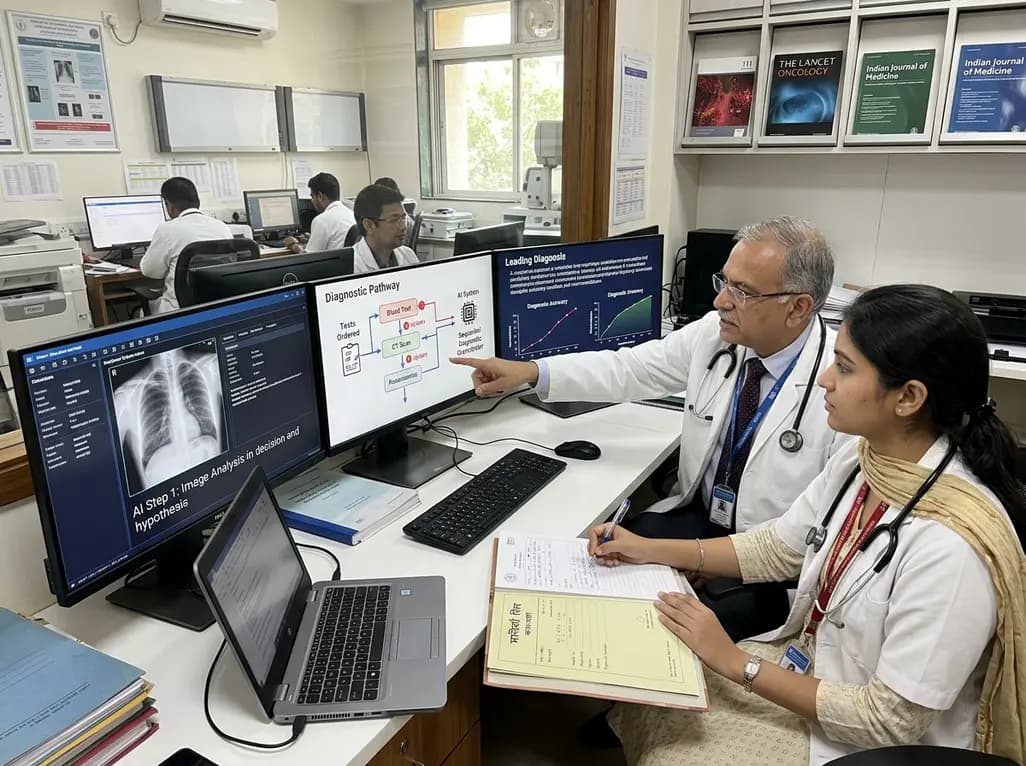
A breakthrough peer-reviewed study demonstrates that a sequential AI diagnosis system can mirror real-world clinical reasoning. By ordering stepwise tests and updating hypotheses dynamically, this multi-agent orchestrator achieved superior diagnostic accuracy while reducing investigative costs.
Today

A prenatal case highlights severe glutaric acidemia type II presenting as bilateral hyperechoic cystic kidneys at 18 weeks. Whole-exome sequencing revealed a novel homozygous ETFDH frameshift variant, demonstrating the power of rapid genetic testing in fetal renal anomalies.
Today

Review how tailored exercise programs benefit frail older patients with heart failure. Explore pathophysiological mechanisms, individualized training prescriptions, and strategies to overcome adherence barriers in clinical practice.
Yesterday

This comparative study evaluates posterior labral tears with and without glenohumeral instability, highlighting distinct demographic patterns, MRI underestimation in stable shoulders, and successful arthroscopic repair outcomes.
Yesterday

A clinical study demonstrates that early intervention with tocilizumab, either as monotherapy or bridged with corticosteroids, achieves significant proptosis reduction and resolves active inflammation in treatment-naive thyroid eye disease.
Yesterday
