.webp&w=384&q=75)

Loading, please wait...
Loading, please wait...
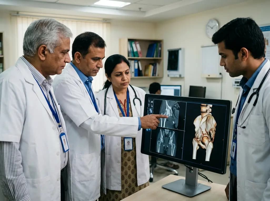
Acetabular superior wall fractures represent a unique and often underrecognized clinical entity in the field of orthopedic trauma. Traditionally, surgeons classified acetabular injuries into ten specific patterns described by Letournel and Judet. However, researchers have identified a transitional grey-zone that exists between the classical trapezoidal anterior wall and large-fragment posterior wall patterns. Specifically, Acetabular superior wall fractures involve the superior acetabular dome, which serves as the primary weight-bearing surface of the hip joint. Because this area is vital for joint stability and mobility, even minor disruptions can lead to significant functional impairment. Surgeons often encounter these fractures in geriatric populations, although high-energy trauma can also cause them in younger adults. Consequently, recognizing these as a distinct fracture entity is essential for improving surgical planning and patient outcomes. Understanding the unique morphology of these injuries allows clinicians to move beyond traditional classification systems that might overlook the complexities of the superior dome. Ultimately, early identification and specialized management strategies are necessary to preserve the longevity of the hip joint.
The etiology of these fractures varies significantly across different patient demographics and energy levels. In younger individuals, high-energy mechanisms like motor vehicle collisions or falls from significant heights typically cause these injuries. During these events, the force vector often drives the femoral head superiorly against the acetabular roof. Consequently, this high-velocity impact frequently results in significant comminution and associated pelvic ring injuries. In contrast, geriatric patients often sustain these fractures through low-energy mechanisms, such as simple falls from a standing position. Because elderly patients often suffer from reduced bone mineral density, their acetabular dome is more susceptible to failure even under minimal stress. Moreover, these injuries frequently involve substantial articular damage to both the acetabulum and the femoral head. Notably, hip subluxation or dislocation occurs in many cases, further complicating the clinical picture. Therefore, clinicians must maintain a high index of suspicion when evaluating patients with hip pain following any mechanism of axial loading. By analyzing the force vectors and patient history, surgeons can better anticipate the specific fracture pattern they will encounter in the operating room.
Accurate diagnosis and preoperative planning for
Management of these complex fractures almost always necessitates open reduction and internal fixation to restore joint congruency. Surgeons predominantly utilize anterior approaches, specifically the iliofemoral approach, to gain adequate exposure to the superior acetabular dome. This exposure is vital because it allows direct visualization of the weight-bearing surface and any impacted fragments. Once the surgeon exposes the fracture, they must carefully elevate any impacted articular segments and support them with bone grafts if necessary. Consequently, stabilization often involves a combination of specialized reconstruction plates and lag screws. Because the superior wall must withstand significant axial loads, the fixation must be robust enough to allow for early mobilization. Additionally, surgeons must address any associated femoral head injuries simultaneously to prevent future joint instability. However, achieving an anatomical reduction is often difficult due to the degree of comminution typically seen in this fracture entity. Therefore, meticulous surgical technique and a deep understanding of pelvic anatomy are paramount for success. Moreover, the choice of implants must account for the bone quality, especially in the elderly population where screw purchase may be compromised.
Despite the best efforts of surgical teams, clinical outcomes for these patients are frequently unfavorable compared to other acetabular fracture types. The primary reason for this is the substantial articular damage sustained by the superior weight-bearing zone at the time of injury. Consequently, many patients develop relevant joint degeneration or persistent symptoms even after a seemingly successful anatomical reduction. Notably, the incidence of post-traumatic osteoarthritis is higher in this group due to the initial cartilage trauma. Furthermore, patients may experience long-term pain and limited range of motion, which significantly impacts their quality of life. Because the superior dome is so critical for ambulation, any residual incongruity leads to rapid wear and tear of the hip joint. Therefore, surgeons must counsel patients on the potential need for future total hip arthroplasty as a salvage procedure. Interestingly, the outcome appears to be more related to the initial damage than the quality of the surgical reduction itself in some cases. Nevertheless, achieving the best possible reduction remains the primary goal to delay the onset of debilitating arthritis. Ultimately, these findings underscore the clinical relevance of treating the superior wall as a distinct and high-risk entity.
As the global population ages, the incidence of these fractures in the elderly is expected to rise. Because geriatric patients often have multiple comorbidities and osteoporotic bone, their management requires a multidisciplinary approach. Specifically, surgeons must balance the need for stable fixation with the risks of prolonged operative time and anesthesia. Moreover, the high rate of unfavorable outcomes in this group suggests that primary total hip arthroplasty might be a viable alternative in selected cases. However, fixing the fracture remains the first line of treatment for most active seniors to preserve their natural joint. Future research should focus on developing better classification systems that specifically include these superior wall injuries to guide treatment more effectively. Additionally, improvements in implant technology, such as locking plates designed for the superior dome, may enhance fixation stability. Therefore, staying updated on the latest traumatic research is essential for any clinician dealing with pelvic injuries. By refining our understanding of this transitional grey-zone, we can offer more tailored and effective care to our patients. Ultimately, the goal is to improve the functional survival of the hip joint across all age groups.
Acetabular superior wall fractures are distinct because they occupy a transitional zone between the anterior and posterior walls. Unlike standard patterns, they specifically involve the superior weight-bearing dome of the acetabulum. This location makes them particularly prone to marginal impaction and severe articular damage. Consequently, they do not fit neatly into traditional classifications and require specialized surgical considerations to manage the critical load-bearing area of the hip.
Surgeons typically prefer the iliofemoral approach for managing these fractures. This anterior-based approach provides the best visualization of the superior acetabular dome and the weight-bearing surface. It allows the surgeon to directly address impacted fragments and perform precise internal fixation with plates and screws. Additionally, it facilitates the use of bone grafts to support the articular surface, which is often necessary due to the high degree of comminution.
Outcomes are often poor because the superior dome is the most critical part of the joint for weight distribution. Even if the surgeon restores joint congruence perfectly, the initial high-energy impact often causes irreversible damage to the articular cartilage. This damage leads to a high rate of post-traumatic osteoarthritis and persistent pain. Consequently, many patients eventually require a total hip replacement, especially those who sustained significant comminution or femoral head injuries during the initial trauma.
Disclaimer: This content is for informational and educational purposes only and does not constitute medical advice or a professional relationship. Always seek the advice of your physician or other qualified health provider with any questions you may have regarding a medical condition. Refer to the latest local and national guidelines for clinical practice.
References
Gänsslen A et al. Acetabular superior wall fractures: a distinct acetabular fracture entity. Arch Orthop Trauma Surg. 2026 Jul 09. doi: undefined. PMID: 42426542.
Letournel E, Judet R. Fractures of the Acetabulum. 2nd ed. Springer-Verlag; 1993.
Tile M, Helfet DL, Kellam JF. Fractures of the Pelvis and Acetabulum. 4th ed. Lippincott Williams & Wilkins; 2015.

Read summarized clinical updates, watch expert medical content, and earn CME certifications right from your smartphone.
Open your smartphone camera to scan and install.





Acetabular superior wall fractures represent a unique 'grey-zone' in orthopedic trauma. Often seen in geriatric patients and high-energy trauma, these injuries involve the critical weight-bearing dome, leading to complex surgical challenges and a higher risk of joint degeneration compared to classic wall fractures.
3 months ago
Following Nigeria's first recorded Ebola case linked to regional travel, the Africa CDC has convened an emergency ministerial summit. This alert underscores critical clinical priorities for healthcare professionals, including differentiating early viral infection from malaria, implementing barrier nursing, and triage.
Today

Leucine Rich Bio has introduced South Asia's first comprehensive vaginal microbiome test, BugSpeaks. By profiling microbial dysbiosis, pathogens, and antimicrobial resistance, this next-generation diagnostic platform provides clinicians with deeper insights into recurrent infections, fertility, and reproductive health.
Today

Review how tailored exercise programs benefit frail older patients with heart failure. Explore pathophysiological mechanisms, individualized training prescriptions, and strategies to overcome adherence barriers in clinical practice.
Today

This comparative study evaluates posterior labral tears with and without glenohumeral instability, highlighting distinct demographic patterns, MRI underestimation in stable shoulders, and successful arthroscopic repair outcomes.
Today

A clinical study demonstrates that early intervention with tocilizumab, either as monotherapy or bridged with corticosteroids, achieves significant proptosis reduction and resolves active inflammation in treatment-naive thyroid eye disease.
Today
