

Loading, please wait...
Loading, please wait...

Stroke remains a major cause of permanent disability and death globally. Clinicians traditionally evaluate obesity using body mass index; however, this metric fails to distinguish between lean muscle and central visceral adiposity. Consequently, researchers have focused on more sensitive anthropometric measurements. A recent cross-sectional study evaluated the relationship between A Body Shape Index and stroke prevalence using United States population data. This metric standardizes waist circumference relative to height and weight to assess abdominal fat deposition. By capturing visceral adiposity accurately, this novel tool offers valuable insights for cerebrovascular risk stratification.
Body mass index remains ubiquitous in routine clinical evaluations. However, physicians frequently encounter vascular events in patients with a normal body mass index. This paradox occurs because standard weight measurements overlook anatomical fat distribution. In contrast, visceral adipose tissue surrounding vital internal organs triggers profound metabolic derangements.
To overcome this diagnostic limitation, investigators developed A Body Shape Index by adjusting waist circumference for height and weight. Consequently, this metric quantifies abdominal fat accumulation independently of overall body size. Furthermore, an elevated score reflects visceral fat expansion rather than peripheral subcutaneous tissue or musculoskeletal mass. In clinical practice, recognizing visceral adiposity provides crucial diagnostic value. Visceral fat actively secretes pro-inflammatory cytokines, including interleukin-6 and tumor necrosis factor-alpha. As a result, patients experience chronic vascular endothelial damage long before metabolic abnormalities appear on routine panels. Moreover, visceral adipocytes release free fatty acids directly into portal circulation, precipitating hepatic dyslipidemia and insulin resistance. Therefore, evaluating abdominal morphology allows clinicians to detect occult cerebrovascular vulnerability that traditional metrics reliably overlook.
The nationwide investigation examined cross-sectional data from 40,213 adult participants enrolled across four NHANES cycles between 2003 and 2018. Researchers identified stroke status using verified personal medical history records. Furthermore, the authors utilized multivariable logistic regression models adjusted for demographic characteristics, behavioral factors, and cardiometabolic comorbidities.
The primary statistical models demonstrated a statistically significant association between higher scores and stroke prevalence. Overall, each incremental increase in the anthropometric index was associated with elevated odds of stroke, yielding an adjusted odds ratio of 1.19. Furthermore, participants in the highest quartile faced a 33 percent greater likelihood of stroke compared to those in the lowest quartile. Additionally, restricted cubic spline analysis revealed a striking nonlinear relationship between this anthropometric measure and stroke risk. Specifically, stroke odds escalated sharply once index values exceeded 0.08. Consequently, this pronounced inflection point suggests that surpassing critical visceral fat thresholds accelerates cerebrovascular pathology. Thus, maintaining values below this threshold represents a clinically meaningful preventive milestone.
Subgroup evaluations provided critical insights into how age, biological sex, and race influence this relationship. Notably, age exerted a profound modifying effect on stroke vulnerability. Among individuals aged 40 to 59 years, elevated index scores conferred an odds ratio of 1.38. For adults aged 60 years and older, the risk rose further to an odds ratio of 1.48.
Conversely, participants under 40 years did not show a statistically significant correlation between index values and stroke prevalence. This finding indicates that prolonged exposure to visceral adiposity over decades compounds vascular damage. Furthermore, aging arteries exhibit reduced compliance and impaired endothelial healing, which amplifies metabolic insults. Importantly, these associations remained robust across male and female participants and varied racial groups. Moreover, the observed cerebrovascular risks persisted independently of baseline body mass index categories. Therefore, a patient possessing a normal body mass index yet high central adiposity remains at elevated risk. Consequently, clinicians must recognize that middle-aged and older adults face disproportionate cerebrovascular hazard from abdominal fat accumulation.
Multiple biological mechanisms explain how abdominal visceral fat promotes cerebrovascular disease. Visceral adipose tissue functions as a dynamic endocrine organ that actively drives atherogenesis. Specifically, visceral adipocytes generate reactive oxygen species that reduce endothelial nitric oxide production. Consequently, cerebral resistance vessels experience impaired autoregulation, persistent vasoconstriction, and accelerated arterial stiffening.
Furthermore, chronic visceral inflammation promotes systemic pro-thrombotic states. Enlarged adipocytes synthesize elevated levels of plasminogen activator inhibitor-1 and tissue factor, which disrupt fibrinolytic pathways. As a result, patients exhibit enhanced platelet activation and increased susceptibility to intravascular thrombosis. Simultaneously, adipokine dysregulation interferes with insulin receptor signaling, causing sustained hyperinsulinemia and atherogenic dyslipidemia. Over time, these pathological alterations induce intracranial lipohyalinosis, microaneurysm formation, and carotid plaque instability. Ultimately, this metabolic cascade heightens the risk of both large-vessel ischemic stroke and small-vessel lacunar infarcts. Therefore, abdominal adiposity acts not merely as a passive marker, but as an active driver of cerebrovascular destruction.
These epidemiological findings hold significant relevance for medical practice in India. South Asian populations notoriously demonstrate the thin-fat phenotype, presenting with high central adiposity despite low body mass index values. Consequently, standard diagnostic criteria consistently underestimate cardiovascular and cerebrovascular danger in Indian patients.
Moreover, stroke incidence in India is increasing rapidly, frequently striking patients a decade earlier than Western counterparts. Incorporating central adiposity assessments into routine primary care screenings could transform preventive neurology. Specifically, identifying patients with index scores above 0.08 provides an actionable early warning sign. Accordingly, clinicians should routinely measure waist circumference alongside standard blood pressure and glycemic checks. When clinicians detect excessive central adiposity, they should promptly initiate aggressive preventive therapies. These interventions include structured dietary counseling, progressive aerobic activity, and strict control of cardiometabolic risk factors. Thus, transitioning beyond simple body mass index allows Indian practitioners to detect hidden vascular vulnerability and prevent life-threatening stroke events.
A Body Shape Index measures abdominal adiposity by standardizing waist circumference against height and body weight. While body mass index merely reflects total body mass relative to stature, this index isolates central visceral fat. Consequently, it accurately identifies cardiometabolic and cerebrovascular risk in individuals who maintain normal body weight.
Restricted cubic spline analysis demonstrates a non-linear threshold effect once the index surpasses 0.08. At this level, visceral fat accumulation triggers intense systemic inflammation, severe endothelial dysfunction, and vascular stiffness. Therefore, exceeding this critical cutoff accelerates arterial atherosclerosis and thrombogenesis, leading to a marked surge in stroke incidence.
South Asian individuals frequently present with the thin-fat phenotype, possessing high visceral fat despite normal body mass index values. Clinicians should routinely record waist circumferences and calculate abdominal adiposity metrics. Thus, identifying elevated values enables physicians to implement aggressive cardiovascular risk factor control and personalized lifestyle interventions promptly.
Disclaimer: This content is for informational and educational purposes only. It does not constitute medical advice, diagnosis, or treatment recommendations. Healthcare professionals should exercise their independent clinical judgment when applying this information. Patient care should always be individualized based on clinical presentation, standard medical guidelines, and diagnostic testing. Refer to the latest local and national guidelines for clinical practice.
References

Read summarized clinical updates, watch expert medical content, and earn CME certifications right from your smartphone.
Open your smartphone camera to scan and install.





A cross-sectional NHANES study reveals that elevated A Body Shape Index (ABSI) is significantly associated with higher stroke prevalence, especially among adults aged 40 and older, with a sharp risk surge above 0.08, highlighting the critical role of visceral adiposity beyond traditional BMI.
Today

The 2025 Australian meningococcal surveillance report shows a 13% decline in invasive meningococcal disease notifications to 118 cases. Genogroup B accounted for 82.2% of typed infections, predominantly impacting infants and young adults, highlighting critical vaccination and rapid diagnostic imperatives.
Today

Doctors at AIIMS New Delhi have achieved a historic milestone by performing India's first open prenatal myelomeningocele repair on a 25-week unborn fetus. This landmark multidisciplinary procedure provides vital neuroprotective benefits, lowers hydrocephalus risks, and transforms maternal-fetal medicine nationwide.
Today
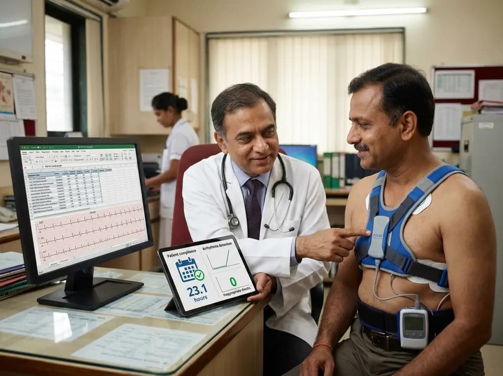
The ACE-PAS registry evaluated 21,612 patients fitted with the ASSURE wearable cardioverter defibrillator. The study demonstrated a 100% ventricular arrhythmia conversion rate, an inappropriate shock incidence of 0.0065 per patient-month, and high daily compliance of 23.1 hours, confirming real-world safety.
Today

A clinical overview of resveratrol supplementation in mitigating exercise-induced muscle damage. This narrative review explores physiological mechanisms, reductions in soreness markers like creatine kinase, dosage strategies, and bioavailability enhancements for sports medicine and nutrition practitioners.
Today

Childhood insulinoma is a rare cause of recurrent hyperinsulinemic hypoglycemia that often eludes early diagnosis. Advanced GLP-1 receptor-targeted PET/CT imaging using 68Ga-exendin-4 complements anatomic MRI to localize occult pancreatic lesions, facilitating focused, curative surgical enucleation.
Today
