.webp&w=384&q=75)

Loading, please wait...
Loading, please wait...
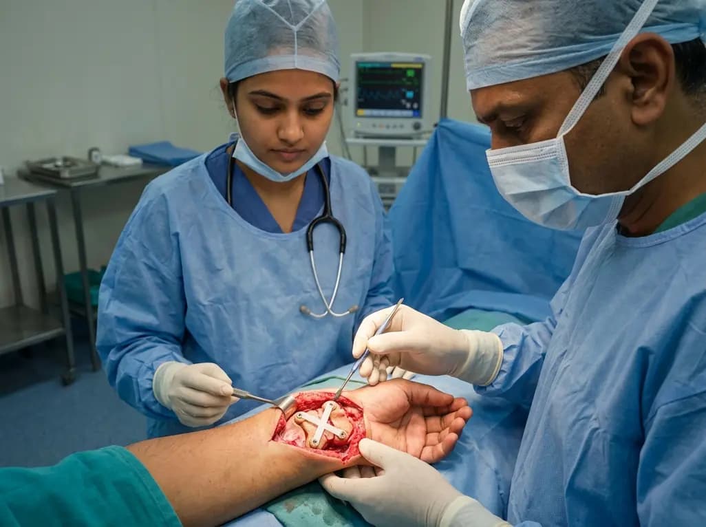
Perilunate dislocations represent high-energy, devastating carpal injuries that often result in long-term morbidity if not managed with absolute precision. These injuries typically involve a sequential disruption of the carpal ligaments, traditionally classified under the Mayfield progression. Historically, surgeons relied heavily on Kirschner wire (K-wire) fixation to maintain carpal alignment during the healing phase. However, the emergence of perilunate injury internal bracing using suture tape technology has recently transformed the surgical landscape. This technique provides immediate mechanical stability while allowing for a more streamlined rehabilitation process. A recent case involving a 31-year-old man highlights the efficacy of this approach. After a high-velocity dirt bike accident, the patient presented with an acute ligamentous perilunate dislocation and significant median nerve paresthesia. Following an initial closed reduction, the surgical team opted for a sophisticated 4-anchor suture tape construct. This modern strategy aims to address the inherent limitations of traditional metal-work by providing a permanent, high-strength internal support system that mimics the natural ligamentous architecture of the wrist.
The stability of the carpus relies on a complex network of intrinsic and extrinsic ligaments, primarily the scapholunate interosseous ligament (SLIL) and the lunotriquetral interosseous ligament (LTIL). During a perilunate injury, the force typically travels through the "lesser arc," disrupting these vital connections in a predictable sequence. Mayfield Stage I begins with the disruption of the SLIL, followed by Stage II involving the lunocapitate joint. Subsequently, Stage III sees the disruption of the LTIL, leading to a perilunate dislocation. Finally, Stage IV results in a complete lunate dislocation into the carpal tunnel. Understanding this progression is essential for effective perilunate injury internal bracing, as the repair must address multiple points of instability. In the discussed case, the patient exhibited a purely ligamentous injury pattern, which is often more challenging to manage than fracture-dislocations. Furthermore, the presence of median nerve symptoms necessitated an urgent carpal tunnel release. By identifying the specific ligaments involved, surgeons can strategically place anchors to recreate the natural biomechanical tension required for carpal harmony and long-term functional success.
For decades, the gold standard for carpal stabilization involved the temporary placement of multiple K-wires across the scapholunate and lunotriquetral joints. While effective at maintaining reduction, K-wires are associated with several significant drawbacks, including pin tract infections, wire migration, and mandatory secondary procedures for removal. Moreover, the prolonged immobilization required with K-wires often leads to permanent wrist stiffness and muscle atrophy. Transitioning to perilunate injury internal bracing addresses these concerns by utilizing ultra-high-molecular-weight polyethylene suture tape. This material offers superior tensile strength compared to traditional sutures and does not require removal. Consequently, the internal brace acts as a "seatbelt," protecting the primary ligament repair during the critical early healing phase. In this 4-anchor technique, the construct is dunked into the carpal bones, creating a stable internal scaffold. This eliminates the need for percutaneous wires, thereby reducing the risk of infection and allowing for an earlier transition to range-of-motion exercises, which is a major advantage for active individuals.
The specific 4-anchor suture tape technique utilized in this case involves a meticulous dorsal carpal approach. The surgeon identifies and reduces the carpal bones before placing four specialized anchors into the scaphoid, lunate, and triquetrum. These anchors serve as the foundation for the suture tape, which is woven to reinforce the SLIL, the LTIL, and the dorsal intercarpal ligament (DICL). Additionally, an extra limb of the suture tape is specifically used to resist excess scaphoid flexion, a common cause of failure in traditional repairs. This comprehensive perilunate injury internal bracing strategy ensures that all three major pillars of carpal stability are addressed simultaneously. Specifically, the construct provides a rigid yet slightly dynamic restraint that prevents the carpal bones from drifting back into a dissociated state. The lack of K-wire retention is a defining feature of this procedure, allowing the surgical team to achieve an anatomical reconstruction that is stable enough to withstand the early phases of physiological loading without the risk of hardware-related complications.
Postoperative management for perilunate injuries has traditionally been conservative due to the fragile nature of ligamentous healing. However, the use of internal bracing allows for a more confident progression through hand therapy. In this case, the patient was immobilized in a short arm cast for a duration of six weeks. Following this period, the patient initiated full range-of-motion exercises under the guidance of a specialized hand therapist. Notably, full weight-bearing and strengthening activities were permitted at the 12-week mark, which is considerably earlier than what is typically allowed with traditional methods. By six months post-surgery, the patient reported minimal pain and achieved a wrist flexion-extension arc of 90 degrees. He was also able to perform a full composite fist and tolerate a dart thrower’s motion, which is essential for daily activities. These results demonstrate that perilunate injury internal bracing not only restores stability but also facilitates a faster return to functional baseline without compromising the integrity of the repair.
The long-term success of carpal reconstruction is often measured by the absence of carpal instability and the patient's reported quality of life. At 32 months postoperatively, the patient in this study demonstrated a Quick Disability of the Arm, Shoulder, and Hand (QuickDASH) score of 14 out of 100, indicating excellent functional recovery. Radiographic evidence confirmed the absence of carpal collapse, fracture, or peri-implant failure, which are common concerns in high-energy trauma cases. Furthermore, the complete resolution of median nerve paresthesia suggests that the initial decompression and subsequent stabilization were highly effective. The successful application of perilunate injury internal bracing in this context highlights its potential as a primary treatment option for complex carpal dislocations. As orthopedic technology continues to evolve, the shift toward internal bracing likely represents the future of ligamentous repair, offering a permanent solution that minimizes the need for secondary surgeries while maximizing the patient's long-term mobility and strength in the wrist joint.
The primary advantage of internal bracing is the elimination of percutaneous hardware and the need for secondary removal surgery. Internal bracing utilizes high-strength suture tape that remains in the body permanently, providing immediate and continuous stability. This approach significantly reduces the risk of pin tract infections and allows for an earlier start to rehabilitation, which typically leads to better range-of-motion outcomes compared to the prolonged stiffness associated with K-wire fixation.
The 4-anchor technique is designed to provide comprehensive carpal stabilization by targeting the major ligamentous complexes. Specifically, an additional limb of the suture tape construct is strategically tensioned to resist excess scaphoid flexion. This is crucial because scaphoid malalignment is a frequent complication in perilunate injuries. By securing the scaphoid to the lunate and triquetrum with multiple anchors, the technique creates a robust internal scaffold that prevents rotational deformities and maintains anatomical alignment throughout the healing process.
Following a 4-anchor internal bracing procedure, patients are usually placed in a short arm cast for about six weeks to allow initial soft tissue healing. After the cast is removed, full range-of-motion and hand therapy begin immediately. By the 12-week mark, patients are typically cleared for full weight-bearing and strengthening exercises. Long-term studies, such as the 32-month follow-up in this case, show that patients can achieve high functional scores and remain virtually pain-free while returning to demanding activities.
Disclaimer: This content is for informational and educational purposes only. It is not intended to be a substitute for professional medical advice, diagnosis, or treatment. Always seek the advice of your physician or other qualified health provider with any questions you may have regarding a medical condition. Refer to the latest local and national guidelines for clinical practice.
References
Hays T et al. A 4-Anchor Suture Tape Internal Bracing Technique for Ligamentous Perilunate Injuries. J Am Acad Orthop Surg Glob Res Rev. 2026 Jul 01. doi: e25.00429. PMID: 42475126.
Mayfield JK, Johnson RP, Kilcoyne RK. Carpal dislocations: pathomechanics and progressive perilunar instability. J Hand Surg Am. 1980;5(3):226-241.
Shin SS, van Eck CF, Papaliodis DN. Suture tape augmentation for the treatment of scapholunate instability. Tech Hand Up Extrem Surg. 2018;22(3):103-107.

Read summarized clinical updates, watch expert medical content, and earn CME certifications right from your smartphone.
Open your smartphone camera to scan and install.





Perilunate dislocations are challenging orthopedic emergencies. This article explores a novel 4-anchor suture tape internal bracing technique that eliminates the need for K-wire retention, facilitating earlier recovery and excellent long-term functional outcomes as demonstrated in a recent clinical case.
3 months ago

Novel diimide and hydrazone scaffolds, particularly compound #C4, synergistically reactivate latent HIV-1 when combined with JQ1 or PKC activators. Demonstrating donor-matched proviral transcription ex vivo and non-canonical signaling, these findings advance shock and kill strategies toward reservoir eradication.
Today

A comparative cohort study evaluates vNOTES vs TLH hysterectomy for benign conditions, demonstrating shorter operative duration, faster return to daily activities, and equivalent female sexual function scores without increased dyspareunia.
Today

Review how tailored exercise programs benefit frail older patients with heart failure. Explore pathophysiological mechanisms, individualized training prescriptions, and strategies to overcome adherence barriers in clinical practice.
Yesterday

This comparative study evaluates posterior labral tears with and without glenohumeral instability, highlighting distinct demographic patterns, MRI underestimation in stable shoulders, and successful arthroscopic repair outcomes.
Yesterday

A clinical study demonstrates that early intervention with tocilizumab, either as monotherapy or bridged with corticosteroids, achieves significant proptosis reduction and resolves active inflammation in treatment-naive thyroid eye disease.
Today
