.webp&w=384&q=75)

Loading, please wait...
Loading, please wait...

3D printing neuroanatomy education is revolutionizing how health professionals master one of the most spatially challenging subjects in medicine. Traditionally, students relied on donor brains or expensive commercial models for learning. However, many institutions globally, including those in India, face significant barriers due to high costs and limited cadaver access. Consequently, additive manufacturing offers a transformative solution by producing high-fidelity, tactile models at a fraction of the price.
One primary advantage of this technology is the ability to visualize complex spatial relationships that 2D diagrams cannot represent. Furthermore, educators can create patient-specific models using actual MRI or CT data. This level of customization allows for a deeper understanding of anatomical variations. Additionally, 3D-printed models are durable and can be handled repeatedly without the fragility associated with cadaveric specimens. As a result, student engagement often increases through interactive, tactile learning experiences.
Adopting this technology requires careful consideration of printing equipment and materials. For instance, resin printers offer superior detail for small neural structures, while plastic filaments are often more cost-effective for larger models. Moreover, sourcing ethically appropriate digital data remains a critical step for educators. Integration into existing curricula should focus on specific pedagogical goals, such as identifying cranial nerve pathways or understanding ventricular systems. Ultimately, these tools bridge the gap between theoretical knowledge and clinical practice.
3D printing allows institutions to manufacture their own anatomical models in-house. This eliminates the need for expensive imported plastic models and reduces the financial burden of maintaining cadaver programs.
Most 3D-printed neuroanatomical models are derived from DICOM files obtained through MRI or CT scans. These files are then processed using specialized software to create printable 3D meshes.
Disclaimer: This content is for informational and educational purposes only. It is not intended as a substitute for professional medical advice, diagnosis, or treatment. Refer to the latest local and national guidelines for clinical practice.
References
Lister JP et al. Next generation open access visualization of neuroanatomy through 3D modeling and 3D printing. Anat Sci Educ. 2026 May 16. doi: 10.1002/ase.70258. PMID: 42141853.
AbouHashem Y et al. The application of 3D printing in anatomy education. Med Educ Online. 2015;20:29847. doi: 10.3402/meo.v20.29847.
Preece D et al. Let's get physical: Advantages of a physical model over 3D computer models and textbooks in learning pelvic anatomy. Anat Sci Educ. 2013;6(4):216-24. doi: 10.1002/ase.1335.

Read summarized clinical updates, watch expert medical content, and earn CME certifications right from your smartphone.
Open your smartphone camera to scan and install.





3D printing offers a cost-effective, high-fidelity solution for neuroanatomy education, overcoming barriers like cadaver scarcity and high model costs....
5 months ago
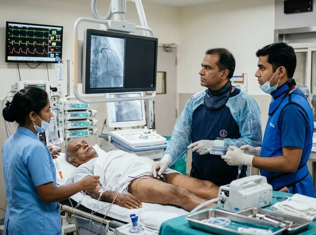
An elderly man with partially repaired Tetralogy of Fallot presented with STEMI caused by coronary embolism rather than atherosclerosis. Successful treatment with sustained mechanical aspiration thrombectomy underscores the vital need for high clinical vigilance in adult congenital heart disease.
Today
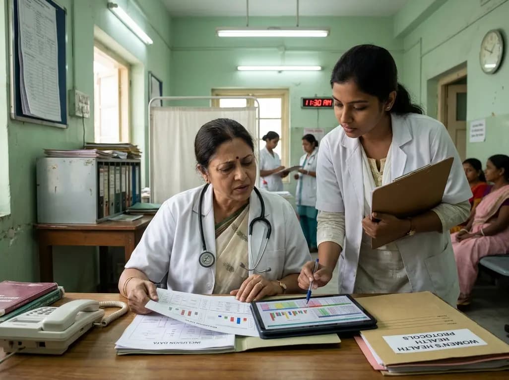
Demanding exhaustive hormonal profiling can inadvertently discourage clinical studies involving females. Methodological standards must align with the specific scientific question to expand, not restrict, equitable evidence for women.
Today
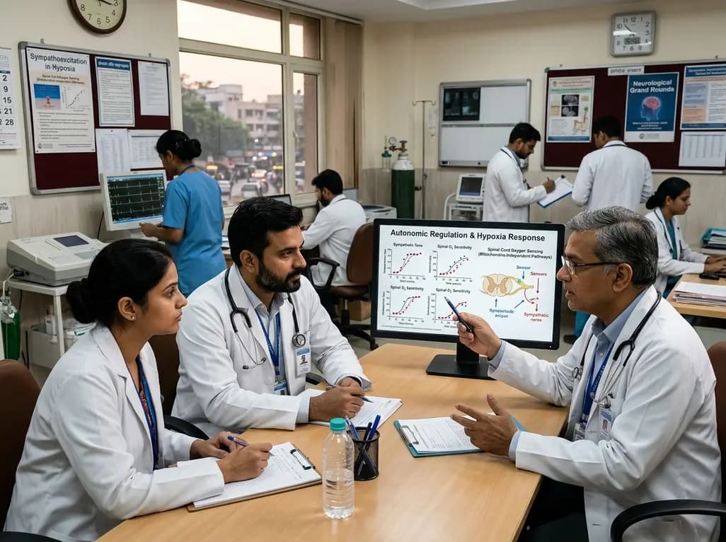
A recent study demonstrates that spinal oxygen sensors do not require mitochondrial electron transport to trigger sympathetic excitation during acute hypoxia, revealing new insights into how mitochondrial oxidative stress modulates autonomic tone in cardiovascular and neurological diseases.
Today
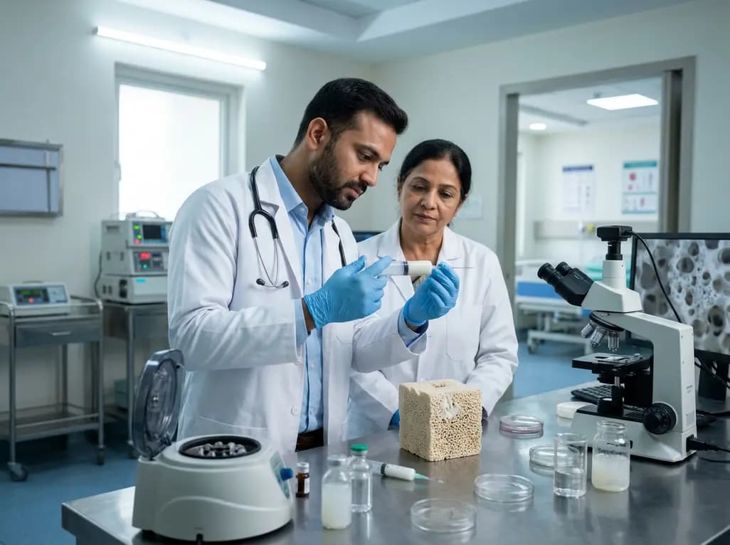
A novel calcium polyphosphate coacervate enables injectable bone implants with in situ hierarchical porosity via phase separation and gas foaming, significantly enhancing cell proliferation, energy metabolism, and osteogenic differentiation for minimally invasive bone defect repair.
Today

Following regulatory scrutiny from FSSAI regarding promotional representations and lean muscle preservation claims, Dr. Reddy's Laboratories and Nestlé Health Science are currently reviewing the branding and marketing strategy for their joint nutritional supplement Celevida GLP+.
Today
