.webp&w=384&q=75)

Loading, please wait...
Loading, please wait...
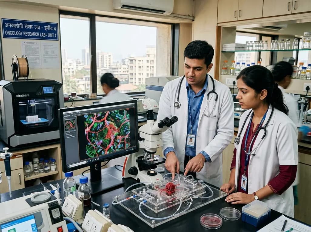
Cancer therapy continues to pose significant therapeutic challenges, primarily because conventional preclinical models fail to replicate human pathophysiology. Traditional two-dimensional cell cultures lack physiological architecture, while animal models frequently display species-specific pharmacological discrepancies. Consequently, bioengineers and oncologists have turned to microfluidic devices that recreate native cellular architecture. Innovative tumor-on-a-chip systems have therefore emerged as transformative platforms for translational cancer research. By integrating additive manufacturing techniques, researchers can now recreate the spatial heterogeneity, mechanical gradients, and vascular networks found within native human neoplasms. These advanced microphysiological devices allow clinicians and researchers to observe therapeutic responses dynamically under controlled fluidic conditions.
Fabricating microphysiological platforms historically required multi-step soft lithography, which proved labor-intensive and structurally restrictive. In contrast, modern additive manufacturing streamlines device prototyping by directly depositing polymeric resins and hydrogels. Stereolithography, digital light processing, and direct extrusion enable investigators to construct intricate microfluidic channels with remarkable spatial resolution. Furthermore, three-dimensional bioprinting permits the simultaneous deposition of living human cells and extracellular matrix components. Hydrogels synthesized from gelatin methacryloyl, alginate, collagen, and decellularized extracellular matrix provide supportive scaffolds that preserve cellular viability during printing. Because printers dispense materials with micrometer accuracy, technicians can engineer delicate perfusion channels alongside parenchymal compartments. Consequently, these automated fabrication techniques ensure batch-to-batch reproducibility across biological testing replicates.
Malignant progression relies heavily on continuous biochemical cross-talk within the surrounding stromal architecture. Accordingly, engineered tumor-on-a-chip systems provide distinct microchambers to co-culture cancer cells with cancer-associated fibroblasts, pericytes, and immune effectors. In addition, integrated microfluidic perfusion channels simulate shear stress and interstitial fluid pressure, which heavily influence neoplastic invasion. Tumors in vivo exhibit steep hypoxia and nutrient gradients that alter phenotypic behavior and promote therapy resistance. Microfluidic designs reproduce these chemical gradients reliably across dense cell matrices. Therefore, researchers can systematically investigate how local tissue hypoxia triggers epithelial-to-mesenchymal transition and extracellular matrix remodeling. By accurately reconstituting these mechanical and cellular dynamics, bioengineered microdevices provide unprecedented insights into invasive malignancy.
Functional vasculature remains essential for sustaining dense cellular aggregates and modeling metastatic intravasation. Fortunately, sacrificial printing techniques allow bioengineers to construct perfusable, endothelialized vascular networks adjacent to solid tumor compartments. Researchers routinely cast pluronic or gelatin sacrificial filaments, surround them with cell-laden bioinks, and subsequently wash them away to leave hollow cylindrical conduits. Endothelial cells rapidly line these hollow lumens, creating biologically responsive vascular barriers under sustained hydrodynamic flow. Consequently, investigators can study how circulating malignant cells penetrate the endothelium and enter the fluid stream. Moreover, adding separate lymphatic drainage channels enables precise quantification of lymphatic dissemination. These vascularized platforms establish a sophisticated experimental testbed for evaluating anti-angiogenic compounds and anti-metastatic therapeutic interventions.
Standard chemotherapy protocols often produce variable clinical responses across diverse patient populations. Patient-derived tumor-on-a-chip systems address this limitation by incorporating primary biopsy specimens into customized fluidic chambers. Clinicians can subject patient-specific microtumors to combinatorial chemotherapy, molecular targeted therapies, and immune checkpoint inhibitors simultaneously. Because these chips replicate physiological perfusion, they reflect drug transport barriers and clearance kinetics far better than static cultures. In neuro-oncology, for example, researchers utilize microfluidic platforms to evaluate therapeutic permeability across an engineered blood-brain barrier toward invasive glioblastoma cells. Similarly, breast and colorectal cancer chips reveal specific resistance mechanisms driven by dense stromal collagen deposition. Hence, personalized microdevices promise to guide patient selection for clinical trials and refine individual treatment regimens.
Despite significant technical milestones, widespread clinical and industrial adoption demands overcoming key practical barriers. Many microfluidic prototypes rely on polydimethylsiloxane, which absorbs hydrophobic drug molecules and biases pharmacokinetic assessments. Transitioning toward inert thermoplastics or specialized bioprintable formulations resolves these drug-absorption artifacts effectively. Additionally, laboratories must standardize sensor integration to enable real-time metabolic and biochemical monitoring without disrupting sterile cultures. Regulatory agencies require robust analytical validation before accepting microphysiological data in place of animal safety studies. Fortunately, active international collaborations are standardizing testing protocols and microfluidic geometries across academic and commercial sectors. As these multi-organ platforms advance, they will accelerate translational discoveries, reduce drug development costs, and refine patient care.
They are advanced microfluidic cell culture devices that simulate the structural, mechanical, and biochemical properties of human malignancies in vitro. By incorporating continuously perfused microchannels, extracellular matrix hydrogels, and multicellular co-cultures, these platforms replicate complex tumor microenvironments far more accurately than standard two-dimensional Petri dishes or non-human animal models.
Additive manufacturing enables rapid, automated fabrication of complex three-dimensional architectures without relying on tedious cleanroom soft lithography. Bioprinting precisely positions multiple cellular phenotypes, structural scaffolds, and sacrificial inks within a single construct. This spatial control allows investigators to generate biomimetic vascular networks, recreate chemical gradients, and ensure reproducible experimental conditions across independent testing devices.
While tumor microdevices cannot completely replace animal models immediately, they significantly reduce reliance on preclinical in vivo testing. These platforms provide highly predictive, human-specific pharmacokinetic and pharmacodynamic data early during preclinical screening. Consequently, researchers can quickly eliminate ineffective or toxic candidates, thereby lowering development costs and accelerating promising oncology compounds toward human clinical trials.
Disclaimer: This content is for informational and educational purposes only and does not constitute medical advice, diagnosis, or treatment. Healthcare professionals must rely on their own independent clinical judgment and institutional protocols when evaluating patient management or diagnostic decisions. Refer to the latest local and national guidelines for clinical practice.
References
Xie Y et al. Developing Tumor-on-a-Chip Systems with 3D Printing Technology. Tissue Eng Part B Rev. 2026 Sep 17. doi: 10.1177/19373368261486383. PMID: 42753260.
Monteiro CF, Deus IA, Custodio CA, et al. Biomaterials meet organ-on-chips: a perspective on tumor modeling. International Materials Reviews. 2024;70(1):31-68.
Zhang B, Korolj A, Lai BFL, Radisic M. Advances in organ-on-a-chip engineering. Nature Reviews Materials. 2018;3(8):257-278.

Read summarized clinical updates, watch expert medical content, and earn CME certifications right from your smartphone.
Open your smartphone camera to scan and install.





Discover how 3D-printed tumor-on-a-chip systems recreate dynamic tumor microenvironments. This technology bridges conventional preclinical models and clinical oncology, advancing precision drug screening, vascular biology analysis, and personalized treatment strategies.
Today

Union Health Minister JP Nadda highlighted patient safety, clinical hygiene, and institutional quality at ABVIMS and Dr RML Hospital on World Patient Safety Day 2026. The national strategy emphasizes NQAS certification, Ayushman Bharat expansions, PMBJP generic access, and safe lifelong management of chronic diseases.
Today

Prelacteal feeding persists in South Asian communities despite known clinical risks. This review examines determinants from Chitral, pathophysiological hazards, and pediatric strategies to eliminate harmful feeds and support exclusive breastfeeding.
Today

A randomized controlled trial demonstrates that theory-driven personalized mobile messaging reduces the risk of consecutive walking goal failures by 33 percent, highlighting the vital role of digital health tools in sustaining long-term exercise habits and chronic disease prevention.
Today

A comparative cohort study demonstrates that patients undergoing total hip arthroplasty after prior hip arthroscopy achieve comparable functional recovery, low complication rates, and equivalent implant survivorship when conversion occurs after six months.
Today

A cross-sectional study of 320 adults evaluated the link between habitual fasting duration and dietary quality using Nutrition Quotient scores, highlighting the clinical necessity of balancing time-restricted eating with nutrient density and meal quality in routine metabolic practice.
Today
