.webp&w=384&q=75)

Loading, please wait...
Loading, please wait...
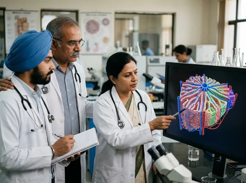
The human liver performs more than five hundred essential biochemical functions while occupying only a modest fraction of total body mass. Interestingly, a single parenchymal cell type—the hepatocyte—executes most of these diverse tasks, ranging from protein synthesis to xenobiotic clearance. Understanding how a single cell type achieves such functional breadth requires exploring the intricate 3D liver architecture. Spatial gradients and microvascular patterns dynamically organize hepatic processes across distinct anatomical zones. Consequently, contemporary research increasingly focuses on structural mapping to decipher hepatic physiology and pathology.
Historically, anatomists proposed multiple complementary conceptual frameworks to explain hepatic microarchitecture. The classical hepatic lobule describes a hexagonal prism organized around a central draining vein. In this framework, portal triads sit at the peripheral vertices, channeling mixed arterial and venous blood through sinusoid networks toward the center. Conversely, the portal lobule model emphasizes exocrine bile drainage centered around a portal bile duct. Furthermore, the metabolic acinus model focuses on unidirectional blood perfusion, dividing parenchyma into three functional zones based on oxygenation, hormone concentrations, and nutrient delivery gradients.
Recent high-resolution serial reconstructions of human liver tissue reveal an even more complex polyhedral modular structure. This modular framework successfully integrates branching microvascular geometry with specialized cellular coordination in three dimensions. Therefore, hepatocytes do not function as isolated biochemical units. Instead, they operate within continuous, polarized three-dimensional cords intimately intertwined with fenestrated sinusoidal capillaries and bile canalicular networks. This intricate spatial arrangement creates localized microenvironments that precisely dictate cellular gene expression and biochemical specialization. As a result, the healthy liver coordinates opposing metabolic pathways simultaneously without cellular competition.
Metabolic zonation represents one of the most remarkable physiological consequences of hepatic microarchitecture. Advanced transcriptomic studies demonstrate that approximately half of all hepatocyte genes exhibit spatially coordinated expression along the porto-central axis. For instance, periportal hepatocytes residing in Zone 1 receive highly oxygenated blood directly from the hepatic artery and nutrient-rich inflow from the portal vein. Consequently, these cells specialize in energy-intensive pathways such as oxidative phosphorylation, gluconeogenesis, fatty acid beta-oxidation, and protective ureagenesis.
In contrast, pericentral hepatocytes located in Zone 3 reside near terminal hepatic venules where oxygen tension declines substantially. These pericentral cells preferentially orchestrate glycolysis, de novo lipogenesis, bile acid synthesis, and cytochrome P450-mediated drug detoxification. Notably, endothelial cells lining the central vein release essential paracrine Wnt ligands, specifically Wnt2 and Wnt9b, along with R-spondin morphogenetic signals. This localized release establishes an instructive molecular gradient that spans the hepatic plate. Consequently, robust beta-catenin activation in pericentral zones drives downstream metabolic programs while suppressing periportal gene expression. Thus, endothelial-parenchymal communication continuously maintains metabolic division of labor throughout homeostasis.
Modern imaging and molecular technologies have revolutionized our ability to dissect hepatic structural organization across multiple biological scales. Historically, researchers relied primarily on two-dimensional histological sections and vascular corrosion casting to infer complex vascular geometry. However, these conventional techniques could not fully capture continuous three-dimensional cellular networks or dynamic spatial gene expression. Today, high-resolution micro-computed tomography provides isotropic, organ-level reconstructions of intricate vascular trees and biliary conduits.
Moreover, optical tissue clearing combined with light-sheet fluorescence microscopy allows intact tissue imaging at single-cell resolution without physical sectioning artifacts. These powerful optical methods preserve three-dimensional spatial coordinates while capturing entire lobular volumes. Simultaneously, single-cell RNA sequencing and spatially resolved transcriptomics have mapped spatial gene expression atlases with unprecedented precision. Techniques such as multiplexed fluorescence in situ hybridization identify distinct transcriptomic shifts across individual hepatocyte layers. Furthermore, multiscale computational models now integrate these imaging and sequencing datasets. These predictive models simulate hepatic hemodynamics, solute transport, and xenobiotic clearance, thereby linking microscopic architecture with whole-organ human physiology.
Because biochemical tasks are strictly compartmentalized along the porto-central axis, hepatic diseases exhibit distinct zonal vulnerabilities. For example, xenobiotic detoxification occurs predominantly in pericentral Zone 3 hepatocytes due to high cytochrome P450 enzyme concentrations. Consequently, acute acetaminophen toxicity causes selective centrilobular necrosis through toxic NAPQI metabolite accumulation and rapid localized glutathione depletion. Similarly, systemic hypotension and hypoxic hepatitis preferentially damage pericentral hepatocytes because baseline oxygen saturation is already lowest near draining central veins.
Conversely, metabolic dysfunction-associated steatotic liver disease typically initiates lipid droplet accumulation in pericentral regions before extending across the lobule. In contrast, toxic insults from heavy metals, iron overload, or viral pathogens frequently provoke prominent periportal injury in Zone 1. Furthermore, hepatic fibrogenesis demonstrates marked spatial heterogeneity, where resident hepatic stellate cells activate in specific microenvironments depending on disease etiology. Toxic and metabolic insults typically trigger pericentral and sinusoidal fibrosis, whereas autoimmune hepatitis and chronic viral infections induce periportal interface hepatitis. Therefore, understanding microarchitectural zonation provides vital diagnostic insights for clinical histopathology and targeted drug development.
The human liver possesses an extraordinary capacity to regenerate and restore both functional mass and complex three-dimensional architecture after acute injury. Following surgical resection or localized necrosis, quiescent mature hepatocytes rapidly re-enter the cell cycle to replenish lost tissue. Interestingly, adult hepatic regeneration recapitulates fundamental embryonic developmental pathways. During this reparative process, central vein endothelial cells dynamically upregulate Wnt ligands and angiocrine factors to orchestrate cellular proliferation and re-establish proper metabolic zonation.
Nevertheless, severe chronic injury and advanced cirrhosis disrupt these delicate regenerative cues, culminating in parenchymal collapse and organ failure. To overcome donor organ shortages, synthetic biology and bioengineering aim to construct transplantable hepatic tissues. Bioengineers currently utilize high-resolution 3D bioprinting, decellularized extracellular matrix scaffolds, and induced pluripotent stem cells to recreate functional tissue units. However, establishing perfusable microvascular networks with durable endothelial-parenchymal crosstalk remains a primary translational hurdle. Successfully integrating spatial morphogen gradients and microfluidic perfusion into engineered constructs will be essential to produce functional liver tissue for clinical therapeutics.
The three-dimensional arrangement of liver lobules directs blood flow sequentially from portal triads to central veins. This directional perfusion generates continuous radial gradients of oxygen, nutrients, and systemic hormones. Simultaneously, central vein endothelial cells secrete morphogens like Wnt and R-spondin. These combined physical and chemical gradients instruct hepatocytes along the porto-central axis to express distinct enzymes, thereby compartmentalizing opposing metabolic pathways across distinct lobular zones.
Because hepatocytes in different zones express distinct metabolic enzymes and experience different oxygen levels, they respond uniquely to stressors. Zone 3 hepatocytes express abundant cytochrome P450 enzymes and experience low baseline oxygen tension, making them highly vulnerable to drug toxicity and ischemia. Conversely, Zone 1 hepatocytes encounter high nutrient and toxin concentrations directly from portal blood, predisposing them to specific infectious agents and metal toxicities.
Recreating functional hepatic tissue requires replicating its complex microvascular geometry and zonal morphogen signaling. Tissue engineers must incorporate perfusable sinusoidal capillaries, functional biliary drainage, and localized biochemical cues into synthetic biomaterials. Without proper three-dimensional organization, cultured hepatocytes rapidly dedifferentiate and lose metabolic specialization. Incorporating spatial Wnt gradients and multicellular co-cultures allows bioengineers to construct mature, biomimetic liver substitutes for translational clinical transplantation.
Disclaimer: This content is for informational and educational purposes only. It is not intended to be a substitute for professional medical advice, diagnosis, or treatment. Always seek the advice of your physician or other qualified health provider with any questions you may have regarding a medical condition. Refer to the latest local and national guidelines for clinical practice.
References

Read summarized clinical updates, watch expert medical content, and earn CME certifications right from your smartphone.
Open your smartphone camera to scan and install.





The liver executes over 500 biochemical tasks through a single parenchymal cell type organized by 3D architecture and morphogen gradients. This review explores structural models, metabolic zonation, imaging advances, and regenerative bioengineering.
last month
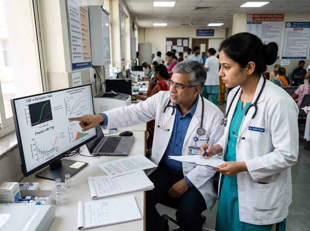
A new PPMI cohort study demonstrates that combining blood microRNA-7-5p with CSF alpha-synuclein seed amplification assay significantly improves the prediction of Parkinson's disease phenoconversion, offering clinicians and researchers a powerful tool for patient risk stratification and clinical trial enrichment.
Today

A large retrospective study evaluates the screening accuracy, positive predictive value, and test failure rates of noninvasive prenatal testing (NIPT) in assisted reproductive technology (ART) pregnancies compared to spontaneous conceptions, highlighting key considerations for pretest counseling.
Today

Differential oxygenation during peripheral TA-NRP can cause undetected coronary hypoxemia due to arch vessel clamping. A novel circuit with continuous aortic root monitoring detects desaturation early, facilitating left ventricular unloading and preventing allograft dysfunction and unnecessary heart discard.
Today

A randomized controlled trial compared ultrasound-guided intra-articular injections of PRP, hydrolyzed type I collagen, and hyaluronic acid in knee osteoarthritis. All therapies demonstrated significant improvements in WOMAC and pain scores at six months, supporting hydrolyzed collagen as a viable biological option.
Today

A scoping review evaluates 116 studies on AI in women's reproductive health, finding that 83.6% carry high risk of bias. While ensemble models show promise, disparities and lack of validation hinder LMIC deployment.
Today
