

Loading, please wait...
Loading, please wait...
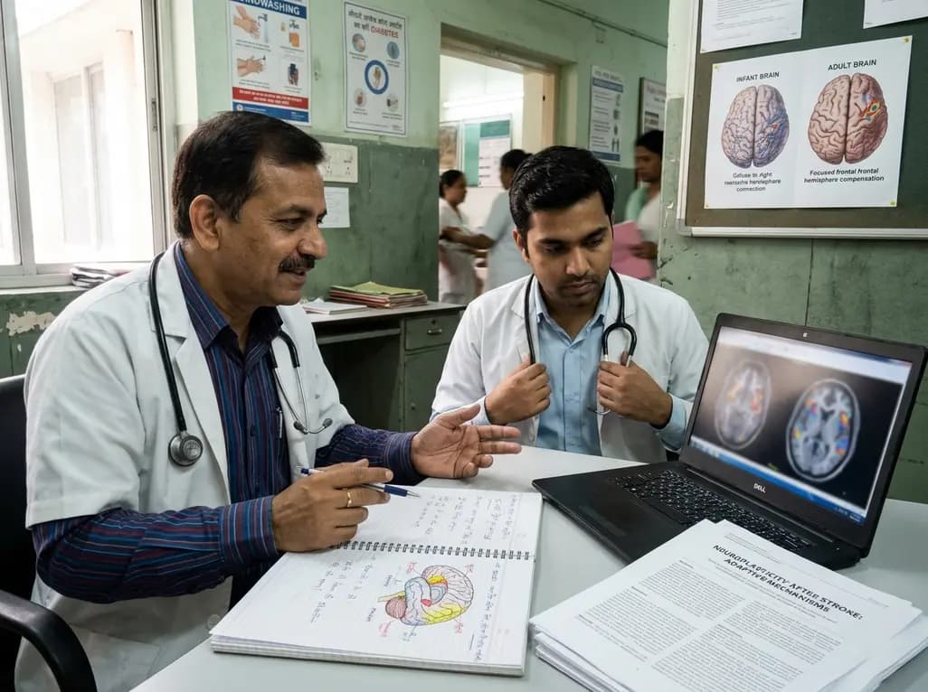
Stroke remains a major cause of acquired neurological disability across India and globally. Consequently, post-stroke aphasia presents substantial clinical challenges for patients and multidisciplinary rehabilitation teams. While traditional aphasiology emphasizes the dominant left perisylvian network, emerging neuroimaging highlights the vital role of right hemisphere language recovery. When cerebral infarction damages classical left-sided language centers, surviving homologous networks frequently engage to re-establish communication pathways. This recruitment demonstrates functional neuroplasticity in real time.
In acute phases, spontaneous upregulation of homotopic frontal and temporal areas occurs across the non-dominant hemisphere. However, clinicians must recognize that compensatory recruitment varies widely across patient cohorts. Early intervention often enhances these spontaneous adjustments, yet maladaptive recruitment can also arise. By understanding how non-dominant structures engage during rehabilitation, neurologists and speech-language pathologists can design personalized therapeutic regimens. Therefore, deciphering these neural dynamics allows clinicians to optimize rehabilitation protocols and guide aphasia recovery effectively.
Perinatal arterial ischemic stroke provides a unique window into developmental neuroplasticity. When severe ischemic injury damages left-sided language zones around birth, the infant brain exhibits profound functional resilience. Remarkably, children who sustain extensive left-hemisphere perinatal infarctions routinely acquire fluent language abilities comparable to healthy peers. Functional magnetic resonance imaging demonstrates that these pediatric patients systematically reorganize core syntactic and semantic networks into homotopic regions of the right hemisphere. Furthermore, even specialized literacy functions relocate successfully to the right fusiform gyrus.
In contrast to adults, pediatric brains accommodate both core linguistic operations and standard right-hemisphere functions within the same hemisphere without overt cognitive crowding. Immature neural substrates retain robust pluripotency and bilateral connectivity. Consequently, young survivors achieve normal communicative fluency, underscoring how age-dependent biological constraints govern neural reorganization. Clinicians evaluating pediatric stroke patients should recognize these positive prognostic capabilities and maintain aggressive supportive rehabilitation therapies.
In contrast to pediatric populations, adult stroke survivors face far greater biological barriers to cortical language remapping. Following an ischemic stroke in adulthood, the left hemisphere remains strongly hardwired for primary phonological and syntactic computation. However, extensive functional neuroimaging reveals that the adult right hemisphere continues to participate actively during chronic recovery. Homotopic regions, including the right inferior frontal gyrus and superior temporal cortex, display marked activation during expressive and receptive tasks.
Moreover, structural studies confirm that increased gray matter density within right posterior temporal regions correlates with meaningful improvements in speech fluency. Nevertheless, debate continues regarding whether this recruitment represents efficient compensation or ineffective transcallosal disinhibition. Some patients exhibit robust bilateral coordination, whereas others suffer from suboptimal processing due to incomplete network integration. Therefore, rehabilitation specialists must carefully differentiate between beneficial right-hemisphere compensation and counterproductive maladaptive interference.
In the healthy adult brain, the right hemisphere specializes in suprasegmental features, non-verbal cues, and emotional prosody. When left-hemisphere stroke forces core language tasks into the right hemisphere, fascinating functional interactions emerge. Neuroscientists frequently examine whether shifting propositional speech into right-sided networks disrupts native prosodic processing. Fortunately, clinical evidence indicates that the right hemisphere can manage emotional intonation and recovered syntactic tasks without inducing mutual interference.
Patients frequently maintain their ability to perceive vocal emotions, interpret sarcasm, and express affective nuance even while engaging non-dominant pathways for communication. Additionally, therapists leverage this preserved right-hemisphere emotional circuitry through innovative clinical interventions such as Melodic Intonation Therapy. By pairing intact melodic perception with linguistic drills, speech pathologists guide stroke survivors toward improved verbal output. Consequently, integrating emotional and melodic processing represents a practical, evidence-based bridge toward comprehensive linguistic recovery.
Translating neuroplasticity research into everyday clinical practice demands a nuanced therapeutic approach. Recently, non-invasive brain stimulation tools, such as transcranial direct current stimulation and repetitive transcranial magnetic stimulation, have gained widespread clinical interest. Physicians frequently apply these neuromodulatory techniques to modulate interhemispheric balance and augment behavioural speech therapy. Specifically, clinicians can apply inhibitory protocols over hyperactive right-sided homologues or apply excitatory stimulation over spared left perisylvian margins.
However, when large left-sided lesions preclude intrinsic ipsilateral recovery, facilitatory stimulation over right-sided language homotopes proves clinically advantageous. Therefore, individualized baseline neuroimaging and precise linguistic profiling remain essential before initiating targeted electrophysiological interventions. In Indian clinical settings, where stroke prevalence continues to increase across diverse patient cohorts, multidisciplinary rehabilitation programs must combine tailored phonological drills with modern neurotechnological tools. Ultimately, bridging neuroimaging discoveries with clinical aphasiology will empower physicians to provide personalized restorative interventions.
Despite substantial advancements in mapping hemispheric recovery, several critical knowledge gaps persist in stroke research. Most clinical studies evaluate small, homogeneous cohorts, which limits the generalizability of neuroimaging findings across broader demographic groups. Furthermore, longitudinal investigations tracking structural and functional reorganization from acute ictus through chronic rehabilitation remain scarce. Researchers still need to delineate the precise temporal windows during which right-hemisphere recruitment exerts optimal therapeutic effects versus when it hampers left-hemisphere restitution.
In addition, future trials must examine how vascular risk profiles, bilingualism, and genetic factors influence neuroplastic potential. In multilingual populations such as India, stroke patients often communicate across multiple languages, creating intricate neural activation patterns that warrant targeted exploration. Addressing these unresolved questions through well-powered, multi-center neuroimaging trials will refine patient stratification. Consequently, identifying precise biomarkers of language reorganization will guide the development of innovative, tailored rehabilitation pathways.
Age at stroke onset serves as a fundamental determinant of post-stroke neural reorganization. In infants and young children, immature cortical networks possess exceptional synaptic malleability, allowing the right hemisphere to assume complete sentence processing seamlessly. Conversely, mature adult brains operate under established lateralization and rigid transcallosal dynamics. Consequently, adult stroke survivors achieve partial homotopic recruitment rather than total bilateral functional reorganization, often requiring intensive speech therapy and neuromodulation to optimize functional communication.
Yes, Melodic Intonation Therapy effectively engages preserved right-hemisphere networks to restore verbal communication in non-fluent aphasia. The right hemisphere natively processes musical pitch, rhythm, and prosodic contours. By translating spoken phrases into melodic, intoned patterns accompanied by rhythmic left-hand tapping, clinicians systematically activate frontotemporal pathways in the non-dominant hemisphere. Consequently, this targeted engagement bypasses damaged left-sided articulatory circuits, enabling stroke patients to regain expressive fluency and spontaneous functional speech.
Non-invasive brain stimulation, including repetitive transcranial magnetic stimulation and transcranial direct current stimulation, selectively modulates cortical excitability during aphasia rehabilitation. In patients with small left-hemisphere lesions, inhibitory protocols suppress maladaptive right-hemisphere activity to reduce interhemispheric competition. Conversely, in patients with extensive left perisylvian destruction, facilitatory stimulation directly enhances right-hemisphere homotopic regions. Therefore, clinicians must carefully evaluate lesion topology and individual functional connectivity before selecting an appropriate neuromodulation strategy.
Disclaimer: This content is for informational and educational purposes only... Refer to the latest local and national guidelines for clinical practice.
References

Read summarized clinical updates, watch expert medical content, and earn CME certifications right from your smartphone.
Open your smartphone camera to scan and install.





New insights into right hemisphere language recovery highlight how early neural plasticity preserves speech after perinatal stroke, while adult stroke survivors demonstrate distinct, adaptive reorganization mechanisms.
5 days back

A nationwide UK Cystic Fibrosis Registry study demonstrates that higher updated mean HbA1c levels correlate with a graded, significant increase in all-cause mortality among individuals with cystic fibrosis-related diabetes, reinforcing the critical need for proactive glycemic monitoring and intervention.
Today

A KGMU study reveals over half of women with breast abnormalities fail to undergo diagnostic confirmation, leading to advanced-stage detection. Discover the socio-cultural barriers, clinical risks of incomplete resection, and community-level solutions needed to bridge the critical screening-to-diagnosis gap in India.
Today

A comparative non-contact study reveals that high-power radiofrequency delivery with the QDOT MICRO catheter induces an artifactual contact force rise. Electrophysiologists must understand this power-dependent thermal and electrical phenomenon to avoid inadvertent catheter withdrawal and ineffective ablation.
Yesterday
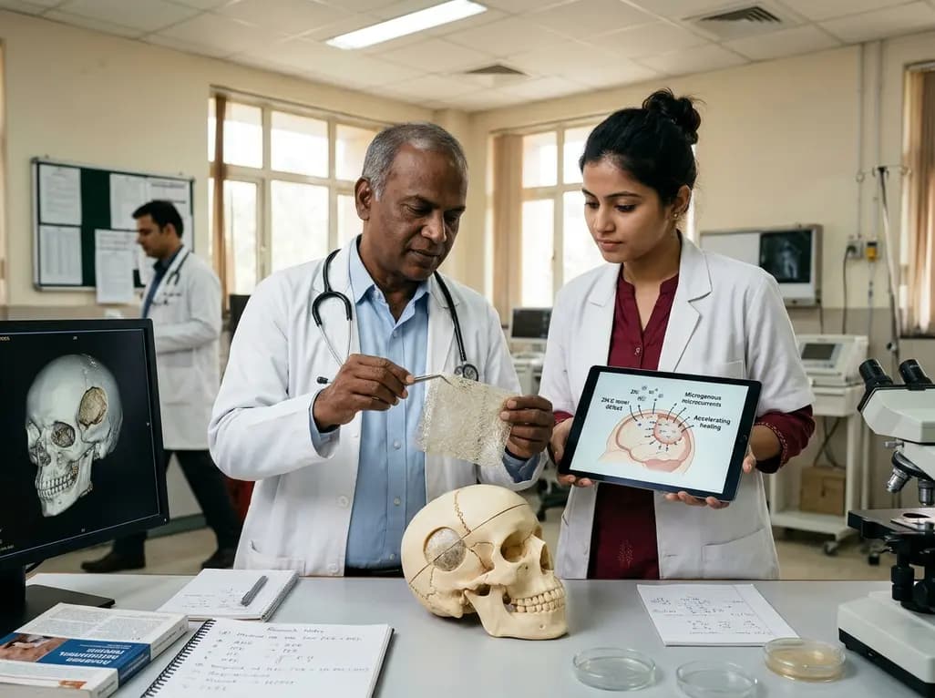
Researchers have engineered a self-powered Zn/C micro-galvanic electrospun membrane that couples sustained zinc release with endogenous microcurrents. This bioelectric scaffold stimulates M2 macrophage polarization and accelerates bone deposition to achieve robust cranial defect regeneration.
Today
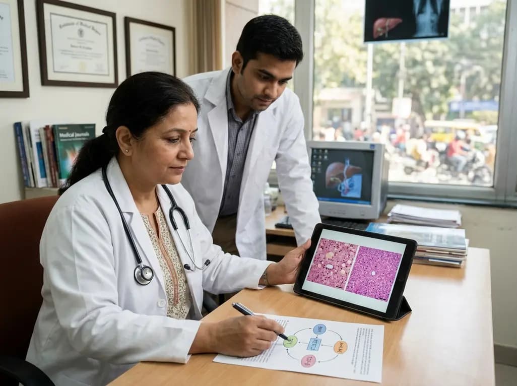
Preclinical research demonstrates that BMI1 overexpression alleviates metabolic syndrome-related liver injury. By triggering ubiquitin-mediated METTL3 degradation, BMI1 prevents AHNAK m6A methylation and suppresses fibrogenic TGF-β/SMAD signaling, significantly improving hepatic steatosis and insulin resistance.
Today
