

Loading, please wait...
Loading, please wait...
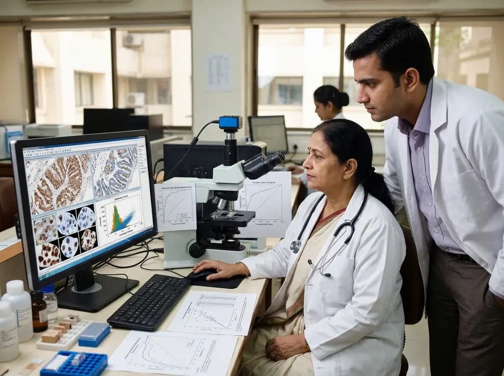
Evaluating p53 immunohistochemistry gastric cancer specimens provides pivotal prognostic insight, yet traditional visual scoring suffers from notable interobserver variability. Because TP53 gene alterations drive significant gastric oncogenesis, pathologists routinely rely on protein expression as a surrogate marker. However, conventional manual assessment struggles with non-standardized threshold cutoffs, which creates significant diagnostic ambiguity. Fortunately, recent developments in computational pathology and deep learning offer automated quantitative workflows that generate continuous nuclear staining metrics across tissue microarrays.
Historically, manual interpretation of p53 staining assigns tumors into categorical tiers, separating normal wild-type patterns from aberrant null or overexpressed phenotypes. While this stratification predicts genetic alterations with moderate accuracy, human visual estimation inevitably introduces subjective bias. Consequently, borderline cases often yield discordant clinical conclusions across independent diagnostic pathology laboratories. To overcome these scoring inconsistencies, researchers have turned to high-resolution whole-slide imaging coupled with artificial intelligence algorithms. Digital pathology platforms now allow investigators to measure cellular staining intensity across thousands of individual neoplastic cells simultaneously. In addition, automated cell segmentation algorithms distinguish malignant nuclei from surrounding non-tumoral stroma with high fidelity. For example, deep learning models such as InstanSeg reliably isolate tumor boundaries without extensive manual intervention. By converting qualitative visual assessments into continuous histochemical scores, digital pathology establishes a stable baseline for comparative biomarker evaluation. Furthermore, automated quantification minimizes labor-intensive slide reviews while improving reproducibility in large patient cohorts.
To quantify nuclear expression with precision, digital image analysis converts raw pixel optical densities into a standardized continuous H-score ranging from 0 to 300. Investigators recently validated this workflow in tissue microarrays comprising 528 gastric adenocarcinoma patients. The computational pipeline detected tumor cell nuclei through specialized neural networks and subsequently measured both staining percentage and optical intensity levels. Crucially, the researchers tested reproducibility across every observer-dependent transition within the digital workflow. Independent manual interpretations achieved 91.7% concordance across tissue cores, establishing a solid baseline agreement. Moreover, the digital H-score demonstrated remarkable algorithmic stability, showing an intraclass correlation coefficient of 0.990 across distinct nuclear detection models. Repeated manual thresholding adjustments similarly yielded an exceptional correlation of 0.954. Thus, continuous digital scoring successfully eliminates the random errors that typically compromise conventional visual inspection. Furthermore, the resulting quantitative scores exhibited a right-skewed distribution, with a median score of 45.5 across all gastric tumor samples.
Although automated scoring provides continuous data, clinical oncology still demands distinct categorical cutoffs to guide patient risk stratification. Therefore, the investigative team evaluated five distinct thresholding strategies to define optimal three-tier classification boundaries. These methodologies included traditional manual thresholds, empirical tertiles, log-rank optimization, the extended Youden index, and volume under the receiver operating characteristic surface. Remarkably, the strategies yielded widely variable lower cutoffs between 8.0 and 27.6, alongside upper thresholds spanning 78.6 to 106.0. The log-rank optimization technique initially produced the widest separation between survival curves. However, its statistical significance vanished completely after rigorous adjustment for multiple hypothesis testing. In contrast, the extended Youden index and volume under the surface criteria produced virtually identical analytical cutoffs. Furthermore, these two multi-class receiver operating strategies maintained strong mathematical convergence across the entire dataset. Consequently, the extended Youden index emerged as a robust, computationally efficient default standard for establishing three-tier p53 categories.
Establishing accurate biomarker thresholds directly influences clinical decision-making and patient survival predictions. In the evaluated gastric cancer cohort, all digital thresholding models successfully separated overall survival curves according to p53 status. Specifically, patients harboring aberrant expression patterns experienced significantly worse survival compared to those displaying physiological wild-type expression. Interestingly, both complete protein loss and extreme overexpression produced comparably poor clinical outcomes. This bimodal hazard pattern aligns precisely with the known biology of TP53 gene inactivation. Null phenotypes typically result from nonsense or frameshift deletions, whereas intense overexpression stems from stabilizing missense mutations. Therefore, precise identification of both aberrant extremes remains clinically vital. Moreover, standardized digital scoring helps identify borderline cases that manual microscopy might misclassify. When digital thresholds clearly delineate wild-type baselines from mutational patterns, oncologists can stratify recurrence risks more confidently. Consequently, automated biomarker quantification can strengthen prognostic nomograms and inform adjuvant treatment selections in routine clinical oncology.
While continuous image analysis represents a major diagnostic advance, several hurdles must be addressed before routine hospital deployment. Most importantly, pathologists must correlate digital H-score thresholds directly with next-generation sequencing data to confirm TP53 mutational status. Although the extended Youden index accurately mirrors manual three-tier groupings, genetic sequencing remains the gold standard for validating true molecular alterations. Additionally, tissue microarrays may not fully capture the profound intratumoral heterogeneity characteristic of advanced gastric carcinomas. Therefore, researchers must validate these digital thresholds across large, whole-tissue resection sections from diverse patient populations. Cross-platform validation across different whole-slide scanners and staining reagents is also essential. Looking forward, artificial intelligence will likely combine p53 quantification with other key gastric biomarkers, such as HER2 and Claudin 18.2. Furthermore, integrating standardized digital metrics into clinical trial workflows will accelerate biomarker discovery and targeted therapeutic development worldwide.
Manual scoring relies on subjective visual estimations of staining intensity and cell percentages, leading to significant interobserver variability. In contrast, digital quantification utilizes deep learning models to measure exact optical densities and nuclear borders. This computational approach yields highly reproducible, continuous H-scores that eliminate human estimation errors, establishing consistent biomarker evaluations across pathology laboratories.
The extended Youden index mathematically balances diagnostic sensitivity and specificity across three distinct phenotypic tiers simultaneously. By using manual expert classification as an objective reference standard, this technique establishes statistically sound cutoffs without overfitting survival data. Consequently, it prevents false statistical significance while matching complex multi-dimensional receiver operating surfaces with greater computational simplicity.
Patients displaying aberrant p53 expression experience significantly worse overall survival compared to those exhibiting normal wild-type expression patterns. Notably, both extreme overexpression caused by missense mutations and complete loss of staining driven by null mutations produce similarly unfavorable prognoses. Accurate cutoff derivation ensures that clinicians correctly identify both aberrant groups to assess individual patient recurrence risks.
Disclaimer: This content is for informational and educational purposes only and should not replace independent clinical judgment. Refer to the latest local and national guidelines for clinical practice.
References
Ahn J et al. Digital image analysis-based quantification of p53 immunohistochemistry in gastric cancer: a systematic comparison of three-tier cutoff-derivation strategies in tissue microarrays. J Pathol Transl Med. 2026 Oct 19. doi: 10.4132/jptm.2026.08.13. PMID: 42898046.
Lee HS, et al. Prediction of TP53 mutations by p53 immunohistochemistry and their prognostic significance in gastric cancer. J Pathol Transl Med. 2020;54(5):378-386.
Bankhead P, et al. QuPath: Open source software for digital pathology image analysis. Sci Rep. 2017;7(1):16878.

Read summarized clinical updates, watch expert medical content, and earn CME certifications right from your smartphone.
Open your smartphone camera to scan and install.





Digital image analysis refines p53 immunohistochemistry in gastric cancer by offering reproducible continuous H-scores. A systematic comparison of cutoff derivation strategies highlights the extended Youden index as an objective, reliable thresholding tool for clinical biomarker evaluation.
6 days back

A nationwide UK Cystic Fibrosis Registry study demonstrates that higher updated mean HbA1c levels correlate with a graded, significant increase in all-cause mortality among individuals with cystic fibrosis-related diabetes, reinforcing the critical need for proactive glycemic monitoring and intervention.
Today

A KGMU study reveals over half of women with breast abnormalities fail to undergo diagnostic confirmation, leading to advanced-stage detection. Discover the socio-cultural barriers, clinical risks of incomplete resection, and community-level solutions needed to bridge the critical screening-to-diagnosis gap in India.
Today

A comparative non-contact study reveals that high-power radiofrequency delivery with the QDOT MICRO catheter induces an artifactual contact force rise. Electrophysiologists must understand this power-dependent thermal and electrical phenomenon to avoid inadvertent catheter withdrawal and ineffective ablation.
Yesterday

Researchers have engineered a self-powered Zn/C micro-galvanic electrospun membrane that couples sustained zinc release with endogenous microcurrents. This bioelectric scaffold stimulates M2 macrophage polarization and accelerates bone deposition to achieve robust cranial defect regeneration.
Today
Preclinical research demonstrates that BMI1 overexpression alleviates metabolic syndrome-related liver injury. By triggering ubiquitin-mediated METTL3 degradation, BMI1 prevents AHNAK m6A methylation and suppresses fibrogenic TGF-β/SMAD signaling, significantly improving hepatic steatosis and insulin resistance.
Today
